● Even better resolution SE 7nm at 3kV, BSE 10nm at 5kV
● New Gun Bias System and Image Signal Processing allows quick and easy focus adjustment and astigmatism correction
● High speed Automatic Focus Control (AFC) and Auto Brightness & Contrast Control (ABCC) function enable faster and optimized image observation
● The new Ultra Variable-Pressure Detector (UVD)(*) is a highly sensitive detector for low vacuum mode
● The new Live Stereoscopic function(*) allows the observation of Stereo SEM image without need for specimen tilting.
|
Items |
Description |
|
|
Resolution |
SE |
3.0 nm at 30 kV (High Vacuum Mode) |
|
7.0 nm at 3 kV (High Vacuum Mode) |
||
|
BSE |
4.0 nm at 30 kV (Variable Pressure Mode) |
|
|
10.0 nm at 5 kV (High Vacuum Mode) |
||
|
Magnification |
x5 to x300,000(on photo*1) |
|
|
Accelerating Voltage |
0.3 to 30 kV |
|
|
Variable pressure range |
6 to 650 Pa |
|
|
Maximum Specimen Size |
200 mm in diameter |
|
|
Specimen Stage |
X |
0 to 100 mm |
|
Y |
0 to 50 mm |
|
|
Z |
5 to 65 mm |
|
|
R |
360° |
|
|
T |
-20 to 90° |
|
|
Observable area |
130 mm in diameter (with rotation) |
|
|
Maximum Height |
80 mm (WD=10 mm) |
|
|
Stage control |
Computer eucentric 5-axis motorization |
|
|
Electron Optics |
Electron Gun |
Precentered Cartridge Filament |
|
Objective Aperture |
5-position, click stop objective aperture |
|
|
Detectors |
Everhart thornley secondary electron detector |
|
|
Analytical Position |
WD=10 mm |
|
|
Display |
OS |
Windows®7 |
|
Image display mode |
Full screen display (1,280 x 960 pixels ) |
|
|
Evacuation System |
Turbo molecular pump |
261 L/s. x 1 |
|
Rotary pump |
135 L/min.(162 L/min. with 60 Hz) x 1 |
|
*1:at 127 mm × 95 mm (4" × 5"Picture size)
*2:at 345 mm × 259 mm (1,280 × 960 pixels)
Application Data
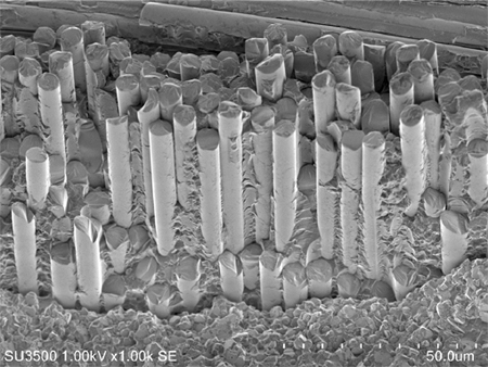
Sample : Filler (Glass fiber) in Resin
Accelerating Voltage : 1.0 kV
Magnification : 1,000x
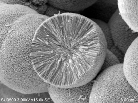
Sample : Titanium Oxide Particle
Accelerating Voltage :3 kV
Magnification : 15,000x
Sample : Courtesy of Prof. Masato Kakihana, Tohoku University
Hiện tại chưa có ý kiến đánh giá nào về sản phẩm. Hãy là người đầu tiên chia sẻ cảm nhận của bạn.