|
Ngày nay, nhu cầu về quan sát và chuẩn đoán bệnh lý ngày càng phức tạp và vượt qua giới hạn quan sát của các loại kính hiển vi quang học (OM). Kính hiển vi quang học có độ phóng đại tối đa đến khoảng 2000 lần nên không thể quan sát được các tổn thương tế bào, mô có cấu trúc siêu nhỏ. Điều này đã dẫn đến quá trình vận dụng kính hiển vi điện tử quét (SEM) để có thể quan sát được các cấu trúc cỡ nanomét trong công tác chuẩn đoán bệnh.
|
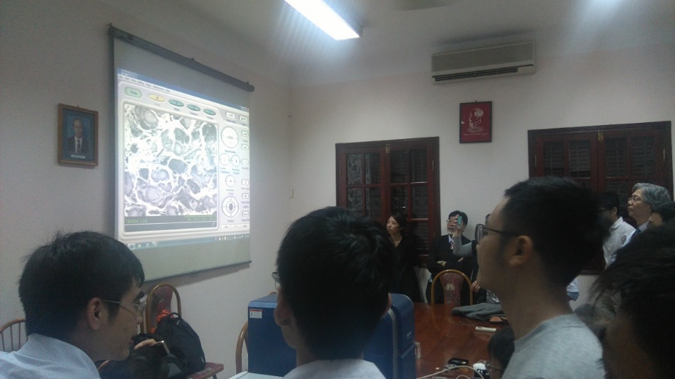
| Loại kính hiển vi điện tử quét (SEM) tiêu chuẩn có chế độ chân không thấp (Low Vacuum) phù hợp cho quan sát các loại mẫu y sinh. Tuy nhiên, thực tiễn sử dụng còn đặt ra yêu cầu là phải dễ sử dụng, kích thước nhỏ gọn, di dộng và đa năng. Loại kính hiển vi điện tử quét để bàn TM3030 của hãng HITACHI là câu trả lời hoàn hảo cho những yêu cầu trên. Trong năm nay, hãng HITACHI đã phối hợp với nhiều bệnh viện đầu ngành tại Việt Nam để tổ chức các cuộc hội thảo chuyên đề về khả năng ứng dụng của kính hiển vi điện tử quét (SEM) trong công tác chuẩn đoán và giải phẫu bệnh phẩm. Các buổi hội thảo này đã được các bác sĩ chuyên khoa quan tâm và đón nhận rộng rãi. Tất cả các chuyên gia đều đồng ý rằng kính hiển vi điện tử quét (SEM) sẽ là công cụ hữu hiệu để quan sát và chuẩn đoán bệnh phẩm yêu cầu độ phóng đại và phân giải cao. Kính hiển vi điện tử quét (SEM) chắc chắn là thiết bị bổ trợ hoàn hảo cho kính hiển vi quang học (OM). Dưới đây là một số hình ảnh về hoạt động hội thảo chuyên đề tại Việt Nam. |
|
|
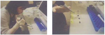
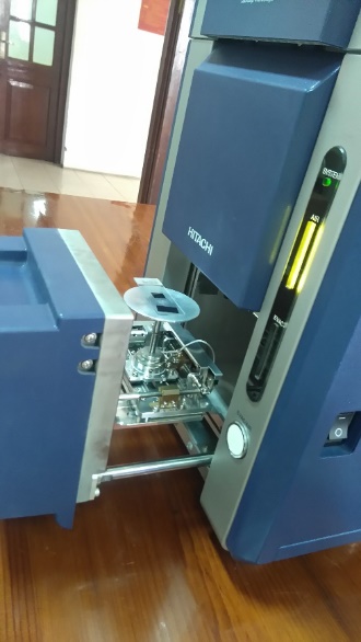
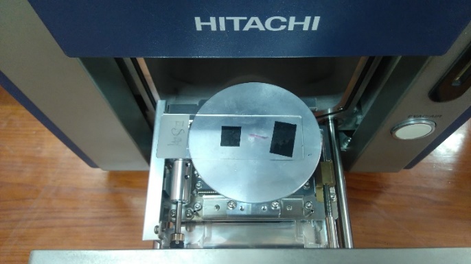
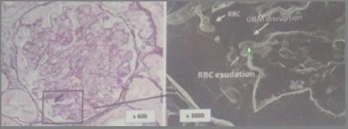
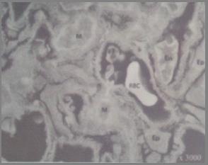
| Mô tả dưới đây thể hiện quy trình chuẩn bị mẫu bệnh phẩm để quan sát dưới kính hiển vi điện tử quét (SEM) và một số kết quả ứng dụng so với kính hiển vi quang học. | |
|
|
|
 |
|
|
|
|
 |
 |
|
|
|
 |
|
|
|
|
 |
|