|
What can we do with the SEM?
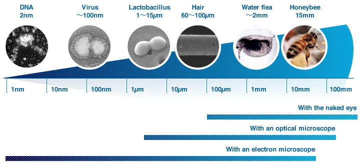
The average human eyes can see objects approximately 0.1 mm in size. To observe objects smaller than this, an optical or electron microscope is necessary.
Electron microscopes are available in a scanning electron type (Scanning Electron Microscope, or SEM) and a transmission electron type (Transmission Electron Microscope, or TEM). The SEM is used particularly for observing the fine structure of a specimen surface at high magnifications, while the TEM is used mainly for observing the inner structure of a specimen at high magnifications. Here, we’ll outline the basic features of the SEM. |
Features of the SEM:
Let's compare images from an optical microscope and those from a SEM.
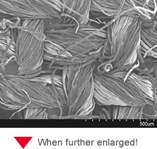
Sample used is a fiber used in parasols for blocking ultraviolet rays.
|
Observation with optical microscope
 |
 |
Fiber observed with an optical microscope. Although the optical microscope provides color information, it has a shallower depth of focus, and when focusing on one particular part of a sample (circled in the image), other parts with different heights become blur.
×110 magnification |
||
|
Observation with SEM |
 |
Fiber observed with a SEM. Although the SEM provides only black & white images (lacks color information), it has a greater depth of focus and provides stereoscopic information.
×110 magnification |
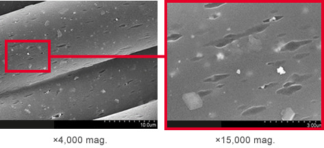 |
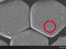
When observing a SEM image by increasing the magnification to ×4,000, inorganic matter (white particles) that are used to block UV rays can be seen scattered inside the fiber. Note that SEM images include secondary electron (shown above) and backscattered electron information (see below). By further increasing magnification to ×15,000, we can observe inorganic matter in the form of particles of 100 to 500 nm in size. A cross-sectional observation reveals how these inorganic particles are dispersed in the fiber.
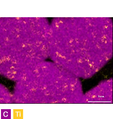
Cross-Sectional Observation and Compositional Analysis of Fiber
 |
Backscattered electron image (×5,000 magnification)
The cross-sectional structure of the fiber was observed with a backscattered electron detector. Now you can see how the white particles are mixed in the fiber.
|
 |
X-ray mapping image of C and Ti/span> |
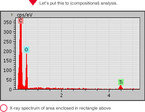
Let's cut the fiber observed above to see its cross-sectional structure by using a backscattered electron (BSE) detector. Since a BSE image helps identify differences in average atomic number as differences in contrast, a location having different compositional elements can be clearly seen. The white glistening particles are inorganic matter scattered in the fiber. When an electron beam is applied to the sample, characteristic X-rays will be produced. By attaching an X-ray analyzer to the electron microscope, you can capture the characteristic X-rays, and learn what elements exist and where they are located.
The spectrum from X-ray analysis on the part enclosed in the rectangle on the BSE imageshows that titanium (Ti) exists in the sample. Now, by conducting X-ray mapping on carbon (C) and Ti in the same visual field as on the BSE image, it will become clear that the fiber contains organic matter (C mainly) and that particles of Ti (actually TiO2), which serve to block UV rays, are dispersed in it.
Principles of the SEM
- Let’s learn the principle of the SEM.-
What is the SEM?
The SEM scans a sample surface with a finely converged electron beam in a vacuum, detects the information (signals) produced from the sample, captures an enlarged image of the sample surface, and displays on the monitor screen.
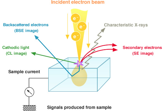
By irradiating the sample with an electron beam in a vacuum, secondary electrons (SE), backscattered electrons (BSE), characteristic X-rays, and other signals are generated as indicated in the figure above. The SEM mainly utilizes secondary electron or backscattered electron signals to form an image. Secondary electrons are produced near the sample surface, and the SE image obtained upon detecting these electrons reflects the fine topographical structure of the sample.
Backscattered electrons are electrons that are reflected upon striking the atoms composing the sample, and the number of these electrons is dependent on the composition (average atomic number, crystal orientation, etc.) of the sample. A BSE image, therefore, reflects the compositional distribution on the sample surface. Also, an X-ray detector can be mounted to the SEM for conducting elemental analysis. So the SEM is usable not only for observing the sample structure, but also as an X-ray analyzer for determining what elements are included in the sample and to what degree.