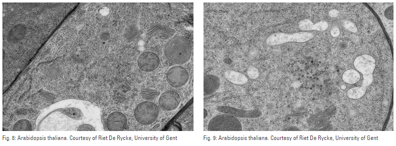
The usual thicknesses for transmission electron microscopic examinations range between 20 nm and 150 nm. There are various techniques for preparing such thin samples. Besides ion etching, FIB, tripod polishing and electrochemical processing, ultramicrotomy is a fast and clean method of producing ultra-thin sections of biological samples as well as polymers, rubber, ductile and even hard and brittle materials. A key advantage of ultramicrotomy is the size and homogeneity of the electron-transparent area of specimens prepared with this technique.
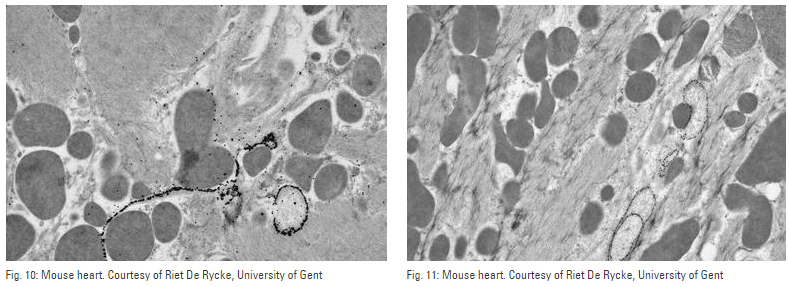
To form an image of a specimen in the transmission electron microscope, electrons have to penetrate the sample without any major loss in speed. A sample’s permeability to electron radiation depends partly on its mass, thickness (thickness × density) and partly on the acceleration voltage of the electron microscope. The electrons absorbed by the specimen can cause a build-up of heat and thus the formation of artifacts in the object.
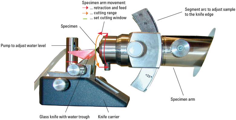
The ultramicrotome is an instrument for producing extremely thin sample sections for examination with an electron microscope. Depending on the kind of sample, it is capable of cutting sections as thin as 10 nm. The sample is inserted into an arm on special bearings that performs a motorized vertical cutting movement. After the section has been cut and the specimen arm retracted, an extremely precise electromechanical feed moves the sample forward slightly by the set section thickness.
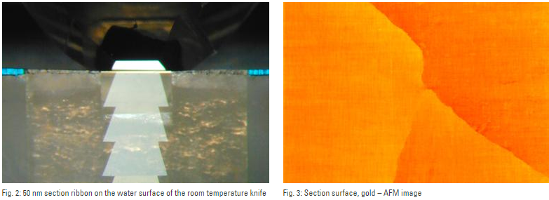
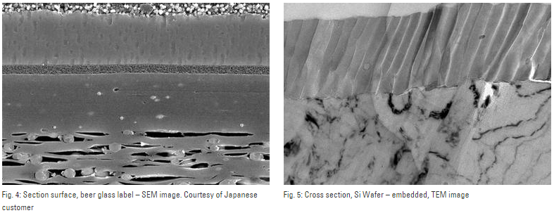
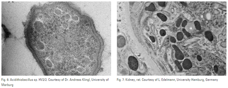
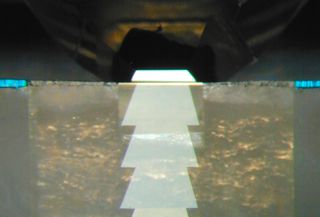
The sectioning is performed by a vertical movement of the sample over the extremely sharp blade of a fixed glass or diamond knife. Removing the sections directly from the knife blade is difficult due to them being so thin. They are therefore collected from the surface of the water bath after the sectioning procedure before preparing them for electron microscopic examination (TEM). In addition the remaining block-face offers a perfectly smooth surface for scanning electron microscopic examination (SEM).