Scanning electron microscope (SEM) is used as a powerful tool to examine the material surface which is wisely applied in Physics and also Life Science. Depending on users' purposes, Sem can be operated to obtain the desired result. In this article, we emphasize the material surface analysys and thickness of material coat measuring by picturing the section.
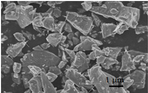
Figure 1: Fe3O4 sample
The photo in SEM, intead of using magnification lenses as optical microscope or transmitted electron microscope, was taken by scanning method. Electron microscope has a suitable depth of focus. The photos of the higher or lower position on the surface are veery clear and have the same clarity at any position (the photo has a good depth of view field). Therefore, using SEM allows for a clearer view of the specimen surface morphology with high resolution and magnification as well as the posibility of calculating the particle size of the material.
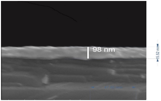
Figure 2: TiO2 sample mixed Nb covered by silver
In the photos of Backscattered Electrons (BSE), the diffences in the average quantity of atoms in the structure will give the different constract, that causes the separation into arrays which is easily recognized in the photos (which element has the higher average quantity of atoms the higher electron backscattering, cause the brighter image and vice versa). Hence by using the section of the material, we could determine the coating thickness or the material thickness by BSE detector. In figure 2, the brighter array is rich silver with the thickness approximately 98nm, the dark array is rich titanium.
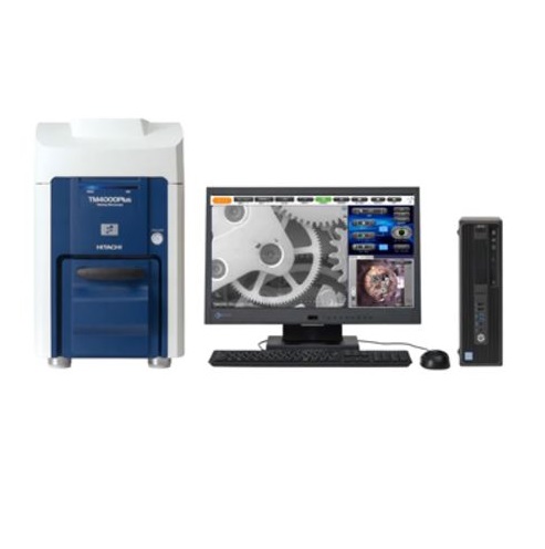
Curently, Red Star Vietnam Co., Ltd announced the grand-opening of the service lab with the function of analyzing surface structure, measuring thickness of coating materials, determining qualitatively and quantitatively the elemental composition of the sample by Scanning Electron Microscopy (SEM) with Energy Dispersive X-Ray Analysis (EDX). The laboratory is equipped with the most modern and innovated instruments using a very powerful specialized software which is operated by our team of genuine trained and professional engineers and technicians, always ready to receive requests and commit to obtain the fastest and most accurate results to our customers.
 |
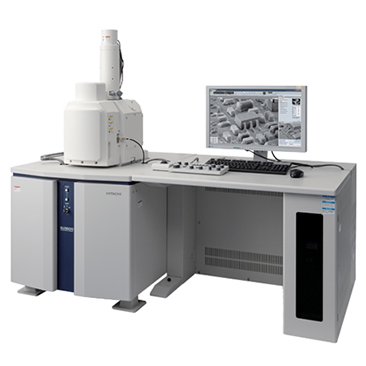 |
Figure 3: Microscopes in demo lab of Red Star Vietnam Ltd.
******************************************
For more infomation, please contact:
Red Star Vietnam Co., Ltd.
Email: info@redstarvietnam.com


