Features
● Ultra-wide field of view, 25X larger volume than comparable systems
● 3 X-ray wavelengths (Cr, Cu and Mo Ka) to optimize imaging for different sample matrices
● Parallel beam geometry for high contrast and rapid data collection
● Auto 5-axis (XYZ and rotation) stage and on-axis imaging system
● High resolution three dimensional (3D) images
● High power rotating anode X-ray source
● High contrast for low-Z materials
● High-resolution CCD imager
In the nano3DX, the magnification takes place in the detector using true microscope elements. This design places the sample close to a high-resolution detector, allowing for a near-parallel beam experiment. This means greater instrument stability and shorter data collection times providing the highest resolution of any X-ray microscope in its class.
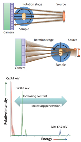
The nano3DX design is a vast improvement over older implementations that use a small source and a long sample-to-detector distance. This geometric magnification requires a very small source and extreme stability to prevent smearing. Data acquisition times can be quite long because small sources are also low power.
The graph at the right shows the three primary anode materials available for use in the nano3DX: chromium, copper and molybdenum, and the effects they have on the experiment. As the energy of the X-ray radiation rises penetration increases but contrast for low atomic weight materials goes down. For bone and silicates, Mo is preferred but for carbon-containing materials, Cu or Cr is preferred. This flexibility is essential to obtaining high-quality, high-contrast images quickly.
Mouse fibula at high resolution
The nano3DX is ideally suited to the analysis of bone samples at submicron resolution. One of the important features of the nano3DX is the ability to look at large samples at high resolution. In the example below, an entire mouse fibula was analyzed with the nano3DX, the first time a whole bone analysis has been performed.
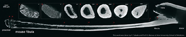
Parallel beam geometry and the ability of the nano3DX to obtain rapid high-contrast images allowed for the collection of the full tomogram of the bone from the proximal to distal ends. With each slice, the structures in the bone are clearly visible, including the softer cartilage at the proximal end. The scale bars are 50 μm long in each of the insets and the bone itself is approximately 12 mm long. The bone marrow, microvessels and osteocyte lacunae are clearly depicted.
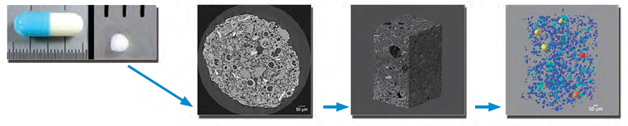
Pharmaceuticals
Below is shown the analysis of a single pharmaceutical particle from a capsule. The first image is a single slice through the reconstruction. The second image is an interior tomogram of the particle at 0.54 μm/voxel. The last image displays a rendering of an analysis of the pores in the sample, color coded by size (red largest, green smallest).
Ultra wide field of view
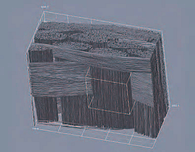
At right is shown an example of a carbon fiber reinforced polymer (CFRP). The fibers are 7 μm in diameter. The image is 1.8 mm x 1.8 mm by 1.4 mm with a voxel size of 0.54 μm. The volume is represented by 3300 x 3300 x 2500 voxels. This volume is 25X larger than the measurable volume from a single scan from other systems at this resolution in a comparable time frame.
High resolution
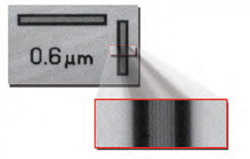
The two dimensional (2D) resolving power is shown directly in the images above with a transmission image of a test pattern at 0.27 μm per pixel in which lines at 0.6 μm are resolved.
| Ống phát tia X-ray | MicroMax-007 HF |
| Điện thế nuôi | 20 to 50 kV |
| Dòng phát | lên tới 30 mA |
| Bia | Cr, Cu và Mo |
| Đầu thu | X-ray CCD camera |
| Số điểm ảnh | 3300 × 2500 |
| Kích thước điểm ảnh | 0.27 to 4 μm |
| Độ rộng trường quan sát | 0.9 × 0.7 mm tới 14 × 10 mm |
| Dải động | 16 bit |
| Bàn mẫu | tự động 5 trục |
| Độ chính xác của bàn mẫu | |
| Độ chính xác của bàn mẫu |
Hiện tại chưa có ý kiến đánh giá nào về sản phẩm. Hãy là người đầu tiên chia sẻ cảm nhận của bạn.