|
The Vertical Turn
Combining Light Sheet and Confocal Leica TCS SP8 DLS
Benefit from two dedicated imaging systems in one: With the Leica TCS SP8 DLS (Digital LightSheet) you get a full confocal with gentle single plane illumination in one microscope. Through the invention of the TwinFlect mirror, we turned our confocal platform Leica TCS SP8 into an easy-to-use, versatile light sheet microscope.
Our turn is THE VERTICAL TURN.
 Liên hệ với chúng tôi Liên hệ với chúng tôi
|
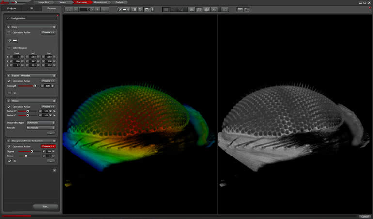
Color coded three-dimensional image volume of eye specific GFP expression (3P3 promoter) in Drosophila melanogaster. Specimen courtesy of Nadja Dinges, Roignant Lab, IMB Mainz |
|
|
Expanded Options
● Increase cell viability with single plane illumination
● Observe fast live processes using an sCMOS-camera
● Enjoy easy sample handling and multi-position experiments
● Discover new fields of applications by combining confocal methods with light sheet microscopy
● Illuminate from two sides to overcome darker regions
● Always find the right imaging method for your application by using the entire world of the Leica TCS SP8
● Turn your confocal into a light sheet instrument. Upgrade from Leica
● TCS SP8 to Leica TCS SP8 DLS at any time
|
|
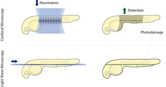
The Vertical Turn – How is Light Sheet brought to a Confocal?
Light sheet microscopy usually requires a dedicated optical setup on an independent system, where the illuminating and detecting objective are perpendicular to each other. The Leica TCS SP8 DLS makes light sheet microscopy as easy as never before. The unique TwinFlect mirror device deflects the illuminating light sheet at a 90° angle and allows the integration of the illumination and detection beam path into the vertical axis of every inverted Leica TCS SP8 without compromising confocal functionality. |
|
Conduct and Document Long-term Observations
Imaging requires light, but too much light can damage your cells. Light sheet microscopy is the most gentle imaging method to date, as it reduces the overall photodamage from phototoxicity and bleaching. This automatically increases the viability of your specimen. Particularly developmental biology benefits from light sheet imaging: The combination of low light illumination and high speed acquisition allows you to follow sensitive developing organisms like a Drosophila embryo over long time periods and to understand how tissue and organs form in real time and 3D. |
Low phototoxicity and specimen imaging in 3D: Development of Drosophila melanogaster over six hours. Probe: Light-sensitive RFP. 3D rendering. 150 µm z stack, 30sec/stack.
|
Principle of Light Sheet Imaging
|
|
Principle of Light Sheet Imaging
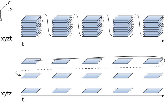
Light sheet microscopy is a highly suitable way of imaging sensitive samples or fast biological processes by illuminating the specimen only in a single plane. Since there is no out-of-focus excitation phototoxic effects can be reduced to the focal plane. It also means that you automatically have optical sectioning and you can image specimens in 3D by moving the sample through the light sheet.
|
Zebrafish Lateral Line Development
|
Multicolor Long-term Observations
17 hours of zebrafish lateral line development. Zebrafish 36hpf, CldnB:lynGFP / Cxcr4B:nuclearRFP. Sample courtesy of Darren Gilmour, EMBL Heidelberg, Germany.
|
|
The Vertical Turn – How is Light Sheet brought to a Confocal?
Light sheet microscopy usually requires a dedicated optical setup on an independent system, where the illuminating and detecting objective are perpendicular to each other. The Leica TCS SP8 DLS makes light sheet microscopy as easy as never before. The unique TwinFlect mirror device deflects the illuminating light sheet at a 90° angle and allows the integration of the illumination and detection beam path into the vertical axis of every inverted Leica TCS SP8 without compromising confocal functionality. |
Principle of Light Sheet Imaging
Light sheet microscopy is a highly suitable way of imaging sensitive samples or fast biological processes by illuminating the specimen only in a single plane. Since there is no out-of-focus excitation phototoxic effects can be reduced to the focal plane. It also means that you automatically have optical sectioning and you can image specimens in 3D by moving the sample through the light sheet.
|
|
Combining imaging modes: Photoconversion (405 nm laser) and wounding (multiphoton laser) in Kaede zebrafish tail. Time lapse recording of photoconverted cell (small red) migrating towards the wound (large red area)
|
Benefit from Many Additional Applications
Our light sheet module is more than a functional add-on to your confocal. The Leica TCS SP8 and Digital LightSheet synergize and give you the possibility to expand your options. You can manipulate specimens using the confocal technology by simply switching between confocal and light sheet mode in LAS X software. Photoconversion or wounding experiments with subsequent gentle long-term observations become easy and convenient. |
Wound Healing by Macrophages
Macrophages in the tail of a 4 day old Zebrafish embryo after wounding (mpeg1:Kaede). Sample courtesy of Francesca Peri, EMBL Heidelberg.
|
|
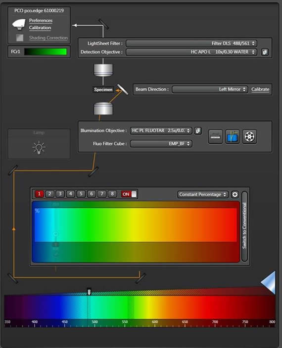
Always the Right Laser
All visible lasers of your Leica TCS SP8 confocal are ready to be used for light sheet imaging. Highest flexibility in choosing the right dye for your light sheet experiment offers the white light laser. It even allows you to use spectrally close fluorophores in multicolor experiments |
|
|
|
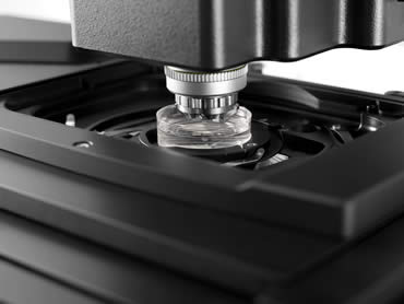
Familiar Sample Preparation
Due to the vertical experimental setup of the Leica TCS SP8 DLS, you can stay close to your familiar sample preparation. The specimens are mounted in conventional glass bottom petri dishes and are directly accessible. You even have the possibility for screening several samples in multi-positioning experiments: Making use of the confocal stage automation, several specimens can be imaged in one experimental setup. The only precondition? Some space for the TwinFlect mirrors on each side of the sample.
|
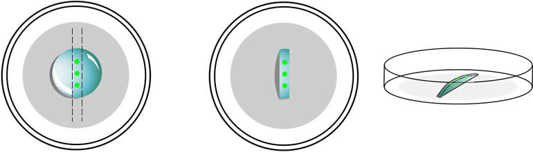
SP8 DLS. Specimens are embedded in agar on glass bottom petri dishes. Excessive agar has to be removed to make room for the TwinFlect mirror.
|
We Focus on Your Needs
Workflow-oriented Software Design
The LAS X microscope software guides users step by step through data recording and evaluation. The workflow-oriented design helps you to use the instrument more efficiently. A convenient calibration routine establishes the light sheet precisely.
Dual-sided illumination of the sample comes by design: Each of the two opposing mirrors of the TwinFlect can be targeted by the scanner to overcome darker areas. For crisp images from a large field of view you can fuse the two images with the online or offline fusion option of the LightSheet Wizard in the LAS X software.
Tailor LAS X to your needs with additional software packages. The LAS X 3D Visualization module provides novel ways to interact with your three dimensional data by intuitive clipping, fast rendering, and stereo display. Tiling experiments enable you to observe large areas, and Mark & Find experiments allow you to observe several regions of interest in a multi-position set up. |
|
Objectives
|
Superior Optics
Superior objectives for a wide range of applications is one of Leica Microsystems’ hallmarks. The heart of our system turning the light sheet vertically is formed by the objectives and the TwinFlect mirror device. With the choice of two different illumination objectives, the Leica HC PL Fluotar 2.5x/0.07 and the Leica HCX PL FLUOTAR 5x/0.15, you can shape the light sheet depending on your experiments’ requirements. In order to reveal finest details or have a larger field of view, you can pick the optimal detection objective, either the Leica HC FLUOTAR L 25x/0.95 W or the Leica HC APO L 10x/0.3 W water dipping objectives. |
|
Convenient Data Handling
Observing processes in 3D and over long time periods generates a lot of data. Several tools implemented into the LAS X software help you to manage your data conveniently. The online fusion tool gives you the choice to keep raw data or to save disc space by saving only the fused image. Your data is automatically saved during acquisition, and the smart loading facilitates data review giving you direct access to the time points or z stacks of interest in large time-lapse data sets. You can compile your desired post-processing steps in a pipeline, which is automatically processed. |
|
Software-Controlled Climate Chamber
A software controlled climate chamber keeps the environmental conditions that your specimens like best. Users gain full control of the experimental conditions with the LAS X Environmental Control module. The logged environmental data can be monitored during the experiment. All environmental conditions can be set within one interface, and you can run temperature profiles, for example, for heat shock experiments. |
|
Hiện tại chưa có ý kiến đánh giá nào về sản phẩm. Hãy là người đầu tiên chia sẻ cảm nhận của bạn.


 info@redstarvietnam.com