|
Today, the requirement of observation and diagnosis of pathology is becoming more complex and exceed the limit of optical microscopes (OM). The magnification of optical microscope is about 2000 times so can't support the observation the cell damage and ultra-small structure tissues. This leads to the operation of scanning electron microscope to observing nanometer structure was applied in the diagnosis.
|
|
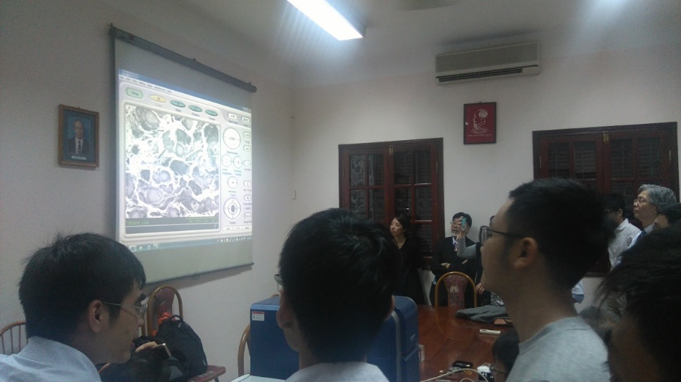
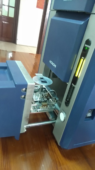
Standard Scanning Electron Microscope (SEM) has the low vacuum mode suitable for the observation of biomedical specimens. However, the practical operation require an instrument easy to use, compact, portable and multifunction. Those requiments can be easily meet with the Tabletop Microscope TM3030. This year, HITACHI has collaborated with many hospitals in Vietnam to organize seminars on the applicability of Scanning Electron Microscope (SEM) in the diagnosis and specimen anatomy. The seminars had been interested and welcomed by top doctors of their specialists. All of thems had agreed that Scanning Electron Microscope (SEM) would be an efficient tool to observe and diagnosis the specimens under the high magnification and resolution. Scanning Electron Microscope (SEM) certainly is a perfect supplementary for optical microscope (OM). Here are some photos of the seminar in Vietnam. |
|
|
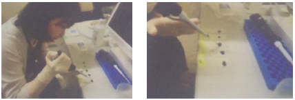
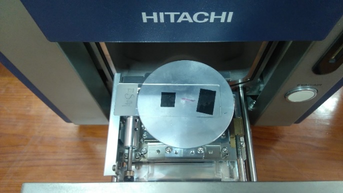
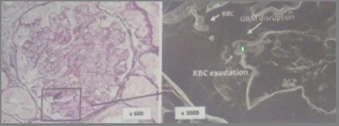
| The description below shows the procedure for preparing specimens for observation under the Scanning Electron Microscope (SEM) and some application results compared to optical microscopes. | |
|
|
|
 |
|
|
|
|
 |
 |
|
|
|
 |
|
|
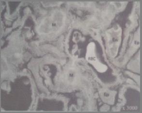
Below is the image of platelet samples taken with a Low-Vacuum Scanning Electron Microscope (LV-SEM). The sample was stained with Platinum-blue (Pt-Blue) according to Inaga method. Thin slices are dyed with Pt-Blue for 20 minutes. It was then observed with the LV-SEM and the cell structure of the specimen can be clearly seen as follows: |
|
 |
|