|
Discover Greater Depths
Multiphoton Microscope Leica TCS SP8 MP
A confocal microscope based on visible light can leave many details invisible. Sometimes the image you need is hidden deep within tissues, which not only scatter light but are damaged by it too.
The excellent optical design of Leica TCS SP8 multiphoton (MP) system makes use of long wavelengths of fully integrated infrared (IR) excitation lasers, the high efficiency of HyD detectors, and an infrared-optimized optical transmission. These allow you to deeply penetrate into the tissue with super-sensitivity and to uncover the finest details of cellular and subcellular processes.
So whether you work with thick organ sections, intact organs or entire live model organisms, let us show you everything the Leica TCS SP8 MP has to offer
 Liên hệ với chúng tôi Liên hệ với chúng tôi
|
|
|
Obtain Excellent Results with Every Sample
● High variety of IR laser sources, with a range of up to 1300 nm ● Fully integrated software control of IR laser ● Superior sensitivity with Leica HyD NDD detectors ● Quad detection module for fast and sensitive multicolor imaging ● High-speed 8 and 12 kHz resonant scanner for fast live cell imaging and fast z stacking ● Superior motCORR IR objectives with motorized correction collar adapt to sample depth ● Dedicated CLARITY and BABB objectives for whole organ imaging up to 6 mm depth ● Modular Leica TCS SP8 platform easily upgrades to MP or other advanced imaging systems (WLL, Fluorescence Lifetime Measurement">FLIM Single Molecule Detection">SMD Stimulated Emission Depletion">STED ● Modular Leica TCS SP8 platform easily upgrades to MP or other advanced imaging systems (WLL, Fluorescence Lifetime Measurement">FLIM Single Molecule Detection">SMD Stimulated Emission Depletion">STED ● Intuitive software control by LAS X For technical specification please click here. |
|
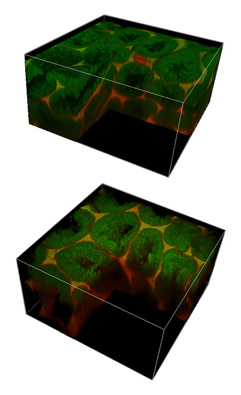
Imagine Imaging More Deeply
The thicker the sample, the more deeply light must penetrate. The more scattering occurs, the less information is obtained.
With multiphoton microscopy, you can use the fact that scattering decreases with longer wavelengths to your advantage. Using light in the red and extended IR spectrum ensures limited scattering and deeper penetration.
Offering motorized correction, dedicated IRAPO objectives and super-sensitive HyD detectors, the Leica TCS SP8 MP optimizes three crucial parameters: optical properties, color correction, and sensitivity.
Make a Deep Impact with the Leica TCS SP8 MP
● Achieve best optical performance with the motCORR motorized correction collar ● Efficiently collect photons with super-sensitive non-descanned HyD detectors ● Optimally correct colors up to 1300 nm with Leica IRAPO high-NA objectives ● Discover broadband anti-reflective coatings for highest transmission in the visible and infrared spectrum |
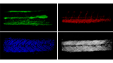
In vivo Thy1-EYFP mouse, z stack. Adult Thy1-EYFP H line mouse, in vivo (cranial window), 800 µm z stack. Courtesy of Dr. Masahiro Fukuda, Department of Molecular Therapy, National Institute of Neuroscience, National Center of Neurology and Psychiatry, Kodaira, Tokyo, Japan.
|
|
|
|
|
|
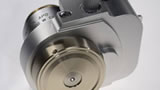
Everything Adapted
The Leica motCORR Motorized Correction Collar
The refractive index (RI) of living cells and their culture medium is close to that of water, making a water immersion objective the best choice. Such objectives are sensitive to RI mismatches caused by variations in coverglass thickness, temperature changes and specimen inhomogeneity. The result is a blurred image and loss of detail in deep tissue.
The Leica motCORR ensures everything is correct and stays corrected. Its motorized correction collar provides an easy, and precise real-time adjustment of the optics to restore optimal imaging in x, y and z dimensions.
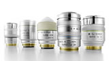
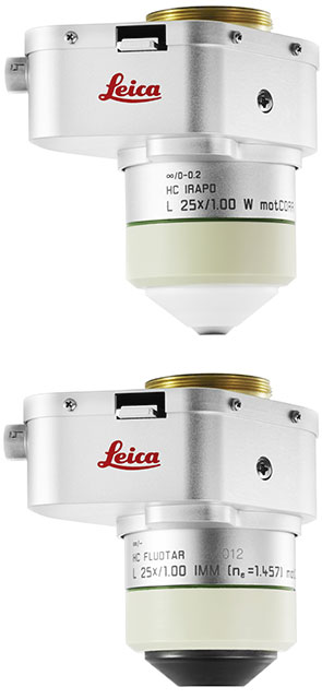
Discover the Leica motCORR Objectives
● Leica HC IRAPO L 25x/1.00 W motCORR − optimized for multiphoton imaging
VISIR − optimized for CLARITY treated specimens
|
|
|
|
|
|
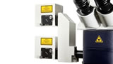
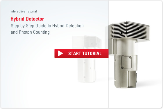
Everything DetectedThe Leica QUAD Module
The great depths you need for whole organ or live cell imaging make detecting very faint structures a challenge. The emitted light is backscattered by surrounding structures, causing the signal to weaken. And simply adding more light can damage the tissue. Combining super-sensitive Leica HyD detectors in a Quad adapter module ensures you can see even the faintest structures in deep tissue sections.
Reach New Depths with the QUAD Module
● Explore deeper tissue sections with a new optical adapter, the Quad module
● Configure the Quad module with up to four super-sensitive Leica HyD detectors in the RLD (Reflected Light Detection) position
● Obtain the highest quantum efficiency of 45 % with the Leica HyD RLDs
● Choose a modular detector set-up (PMT and/ or HyD) and get the super-sensitivity you need with the chance to upgrade later
For technical specification please click here. |
|
|
|
|
|
|
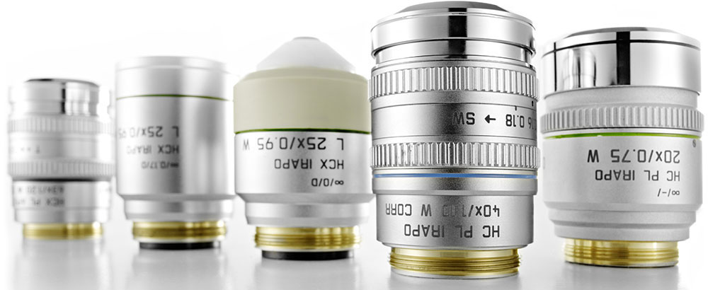
The Correct ApproachImprove MP Imaging with Highly Specialized IRAPO Objectives Understanding the spatial relationship of structures is vital in multicolor specimens. In MP microscopy, infrared objectives require color correction for perfect colocalization in a multicolor sample. Leica IRAPO objectives are specifically designed with perfect color correction and high transmission in mind. They provide bright images while reducing the risk of photodamage by requiring less laser power.
Experience Apochromatic Correction at a New Level
● Achieve excellent multicolor MP imaging by color corrected IR apochromats from at least 700 nm up to 1300 nm ● Maximize the number of photons available for multiphoton excitation and detection with >85% transmission from 470 to 1200 nm ● Experience brighter images ● Reduce photodamage ● Consider the HC IRAPO L 25x/1.00 W motCORR with motorized correction collar for improved MP imaging |
|
An Astonishing Revelation in Live Imaging
High-speed imaging and great image quality: This is the challenge. More light for brilliant imaging means live cells suffer from phototoxic effects and bleaching.
The Leica TCS SP8 MP overcomes this. In MP microscopy, two or more photons reach the fluorophore simultaneously, but only in the focal plane. This reduces out-of-focus excitation compared to confocal microscopy, reduces photodamage, and increases cell viability. In addition, the Leica TCS SP8 MP offers high speed and efficient photon collection to catch the fastest kinetics and the faintest signals.
Discover Our Live Imaging Capabilities
● Use a high speed 12 kHz resonant scanner, offering 428 frames per second ● Enjoy the largest FOV (Field-Of-View) scanner ● Attach up to 4 non-descanned HyD detectors in the Quad module ● Perform electrophysiology and intravital imaging To find more application videos click here. To learn more about the Multiphoton principle click here. |
|
|
|
|
|
|
A Perfect Working Relationship
The Tandem Scanner
With the Tandem Scanner Leica offers the perfect relationship between a large field of view (FOV) and a full range of scan speeds – the ideal combination for live specimen imaging. If your research covers everything from rapid calcium dynamics in brain slices to whole organ samples, the Leica TCS SP8 MP will be in synergy with you.
All in Time
● 22 mm FOV – the largest of any point scanning systems for scanning in one shot with the patented x2y scanning mirror concept
● Unique combination of FOV and 8 kHz or 12 kHz resonant scanning system with switchable galvanometric mirrors
● Scan at frequencies of up to 12 kHz, resulting in about 40 fps (512 x 512 px) or 428 fps (512 x 16 px)
● Acquire 4D stacks up to 50% faster than comparable systems by combining them with the SuperZ GalvoFlow
● No compromise in resolution, sensitivity or contrast, thanks to high-NA objectives and the sensitive Leica HyD RLDs |
|
|
|
|
Everything DetectedThe Leica QUAD Module – Superior Sensitivity
Not only depths, but also speed can make detecting very faint structures in live samples a challenge. If you want to observe fast kinetics, you can increase scan speed, but this means less photons will be collected.
With the Quad module, you can combine up to four super-sensitive Leica HyD detectors to image multiple colors simultaneously. The more sensitive your detector, the brighter your acquired live image. Due to the HyDs´ remarkably low dark noise and their high sensitivity you can reduce laser excitation power, and therefore specimen damage and photobleaching.
Reach New Sensitivity with the QUAD Module
● Explore highest sensitivity with the Leica HyD RLD which offers 45% quantum efficiency
● Reach superior signal-to-noise for brighter images
● Protect your live sample by reducing laser power
● Configure your Quad module with up to four super-sensitive Leica HyD detectors in the RLD (Reflected Light Detection) position |
|
|
|
|
|
|
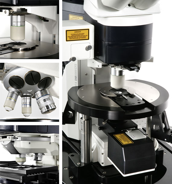
Everything FixedIntravital and Electrophysiological Imaging with the Leica DM6000 CFS Sensitive experiments like patch-clamp electrophysiology require high stability. Even minor movements can distort the imaging results. Core to the Leica TCS SP8 MP platform is the Leica DM6000 CFS microscope. Offering a fixed stage with vibration-free mechanical stability and almost entirely noise-free electronics, it provides an excellent platform for whole-specimen and electrophysiological imaging.
● Switch and dip objectives smoothly without disturbing the experiment using the motorized, remote-controlled 2-fold objective changer
● Perform simultaneous MP imaging and electrophysiological recording due to objectives with inert ceramic fronts and infra-red color correction
● Save time by directly correlating voltage recordings with fluorescence intensity data with the dedicated LAS X electrophysiology software package
|
|
See Color Perception in a New Light
Multiple-labeled fluorescence provides insights into localization and interaction. But imaging multiple fluorophores can result in cross-excitation when laser wavelengths are limited.
With the Leica TCS SP8 MP, cutting-edge infrared lasers offer IR excitation even into the far red. And using multiple hybrid detectors systems, multicolor sensitivity is drastically improved.
Perceive the Benefits of Multicolor Experiments with the Leica TCS SP8 MP
● Use a large variety of IR lasers that are tunable from 680 to 1300 nm ● Discover red and far red dyes with InSight DeepSee+ or OPO MPX ● Combine one or two IR lasers for advanced multicolor imaging ● Achieve simultaneous 4-channel multicolor detection with up to 4 Leica HyDs in a Quad module ● Discover additional label-free information from second (SHG) and third (THG) harmonic generation signals |
|
|
|
|
|
|
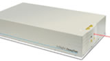
Everything ExcitedAdvanced IR Imaging with the Laser of Your Choice
With the Leica TCS SP8 MP you can make full use of the most advanced infrared lasers on the market and take your multiphoton imaging to new depths. Tune gap-free from 680 nm to 1300 nm through the intuitive LAS X software with a range of lasers that reaches from ultra-fast prechirped femtosecond lasers to the Optical Parametric Oscillator (OPO MPX) and InSight DeepSee+.
Make Exciting Discoveries with the Leica SP8 MP
● Perform gap-free tuning of multiphoton excitation from 680 to 1300 nm
● See deeper and brighter than ever before with higher power, shorter pulse MP imaging
● Extend the IR range with novel red-shifted fluorescent proteins and dyes
● Configure with two MP lasers for the most flexible multicolor excitation
● Conveniently choose wavelengths using intuitive LAS X software
|
|
|
|
|
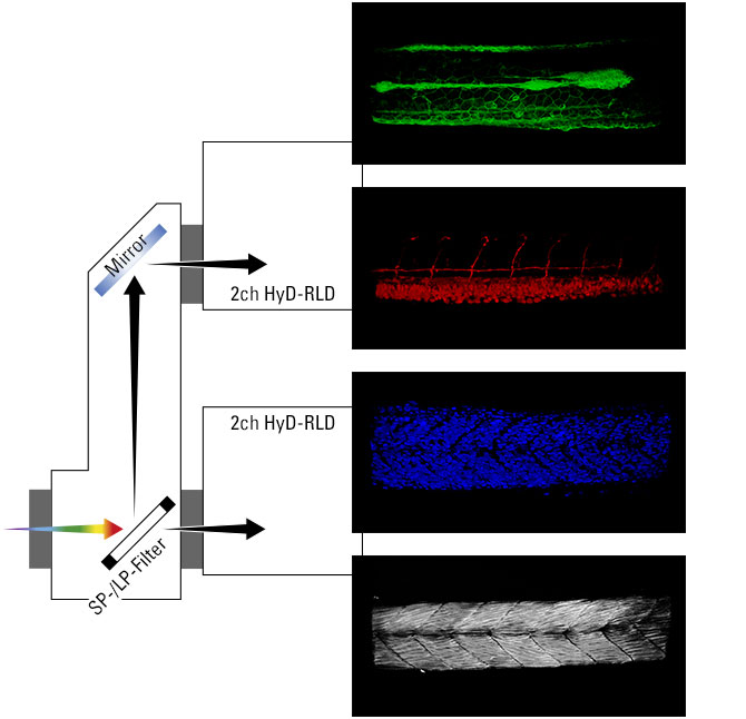
HyD RLD Quad module for four color acquisition.
|
Everything DetectedThe Leica QUAD Module – Faster Multicolor Imaging
Sometimes, observing the localization or dynamics of just one structure provides a limited picture without seeing relationship to its neighbors.
The Quad module improves multicolor super-sensitivity. With up to four Leica HyDs attached, you can fast acquire multicolor and gain insights in localization and interactions from deep within tissues or living specimens.
Reach More Colors with the Quad Module
● Save time by acquiring more colors in one shot
● Observe fast and easy multicolor imaging by combining the Quad module with the high-speed Tandem Scanner
● Configure the Quad module with up to four super-sensitive Leica HyD detectors in the RLD (Reflected Light Detection) position
● Chose a modular detector set up and obtain the highest flexibility for fast and easy multicolor deep tissue acquisitions
|
|
|
|
|
Everything and MoreLabel-free Imaging Through Second and Third Harmonic Generation (SHG/THG)
By employing infrared femtosecond lasers, you can see more than just fluorescence. The high energy produced by these short-pulsed IR lasers is sufficient to create another photoelectric effect without the need for labeling. These are SHG and THG, non-linear polarization effects in the illuminated sample.
SHG signals occur from large centrosymmetric structures with spatial order including collagen, myosin or microtubules. THG can be used to detect an interface between materials of different excitability, for example, the outline of cells in embryos, where the cells are separated by water.
See More with the Leica TCS SP8 MP
● Discover more tissue and live cell imaging without using a dye
● Achieve the brightest SHG and THG imaging with super-sensitive HyDs and dedicated filters
|
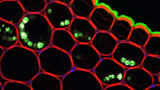
Zebrafish (gastrulation) RNA injection for Neptune fluorescent protein cytoplasmic staining. THG (in blue). 20x1NA; Insight at 1140 nm. Courtesy of Dr. Nadine Peyrieras, BioEmergences, Gif sur Yvettes, France.
|
Thy1-YFP mouse brain treated with CLARITY. Animation generated with LAS AF 3D Visualization. Courtesy of Karl Deisseroth and Raju Tomer, Stanford University, Palo Alto, CA, USA.
|
Let's Get Clarity
Many existing techniques for brain imaging are limited to small regions from thin section, which impedes the study of cellular connection and pathways. With clearing techniques like CLARITY and BABB you can image tissues whole and intact.
The Leica HC Fluotar L 25x/1.0 IMM motCORR VISIR (ne=1.457) is a specialist immersion objective, specifically designed to maximize results from CLARITY-treated specimens. So, by combining CLARITY with the Leica TCS SP8 MP, you can maximize imaging depth and resolution. Click here to see the exciting Nature movie „See-through brains“ from Karl Deisseroth.
Discover perfect CLARITY
● Image whole organs up to a depth of 6 mm
● Match the refractive index of CLARITY-treated specimens for bright, high-resolution images deep within tissues
● Optimize images without disturbing the specimens with a remote-controlled motorized correction collar
● Single- or two-photon excitation possible due to the broad range of color (VISIR) correction |
|
Everything ClearDedicated BABB Objective for Neuroscience and Developmental Biology
Optical performance in neuroscience and developmental biology requires a team that works in perfect synergy. By combining the core microscope platform DM6000 CFS with the Leica HCX APO L20x/0.95 IMM objective and BABB immersion medium you can reveal the secrets of unexplored depths.
● Analyze brain slices, image whole organisms for morphological studies, and discover the anatomy of developing organs
● Optimize your whole mount imaging by using BABB (Benzyl Alcohol Benzyl Benzoate, 1:2)
● Leica HCX APO L20x/0.95 IMM with special front lens suitable for immersion medium BABB ( = 1.559)
● Low magnification 20x at high resolution (NA 0.95)
● Large free working distance of 1.95 mm |
|
|
Shaped for Things to Come
The Leica TCS SP8 MP is configured for your research today but adaptable for tomorrow. Like all Leica Microsystems’ confocal products, the Leica TCS SP8 MP allows you to build and tailor your system upon its easy-to-use core. It can evolve, adapt, and reconfigure to your needs. So, as one discovery leads to another, you can keep the Leica TCS SP8 MP at the very heart of everything you do
Build Upon the Flexible and Easy-to-Use Leica TCS SP8 MP
● Multiphoton and confocal microscopy – two worlds in one system
● Start with the confocal SP8 platform today and adapt it in future with new lasers, new detectors, or a high-speed scanning module
● Tailor it to your discoveries by adding a wide range of modules like SMD, the White Light Laser or super-resolution STED 3X to resolve structures smaller than 50 nm
● Tailor it to your discoveries by adding a wide range of modules like SMD, the White Light Laser or super-resolution STED 3X to resolve structures smaller than 50 nm |
|
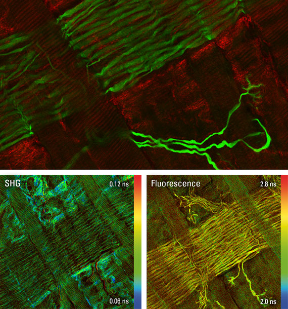
Mouse diaphragm. Signal from muscle (red), motor neurons (green). Courtesy of R. Rudolf, Karlsruhe Institute of Technology, Germany.
|
Adapt it to FLIM
The fluorescence lifetime is a measure of how long a fluorophore remains in its excited state before returning to its ground state. FLIM (Fluorescence Lifetime Imaging) lets you display the fluorophore lifetime in every single pixel, adding an extra dimension to your research.
Using the fast Leica HyD RLD detectors for FLIM-FRET (Fluorescent Resonant Energy Transfer) experiments you can easily explore molecular interactions in depth.
Create High-quality FLIM Data From Large Z Stacks
● Combine femtosecond MP excitation with the non-descanned Leica HyD RLD detectors
● Enjoy a reliable and fast experimental set-up with the dedicated FLIM application wizard
● Be reassured by the user-friendly interaction of Leica and Picoquant components
|
|
Powerful Performance Made SimpleThe Leica Application Suite X
Performing experiments and analyzing your data shouldn’t be as complex as the science you perform. When designing the LAS X, we focused on just two things: powerful features available for microscopy and an intuitive usability from interface to workflow.
Step-by-Step Guidance with LAS X
● Perform your MP experiments efficiently with the help of a workflow-orientated design
● Gain full control of all motorized sliders and IR lasers for fast experiment setups
● Dedicated MP FLIM wizards make data evaluation truly simple
Adapt It to Your Needs
Wherever your multiphoton experiments take you, the LAS X software will provide you with powerful analysis options.
Step-by-Step Guidance with LAS X
● LAS X Electrophysiology
● LAS X 3D Visualization
● LAS X 2D/3D Analysis
● Huygens MP Deconvolution
● LAS X Live Data Mode
● LAS X FLIM Wizard
● LAS X FRET/ FRAP Wizards
|
Hiện tại chưa có ý kiến đánh giá nào về sản phẩm. Hãy là người đầu tiên chia sẻ cảm nhận của bạn.


 info@redstarvietnam.com