The Leica DM2500 LED offers the above mentioned contrasting methods to meet each specimen’s demands:
Especially in the clinical sector, a clear differentiation of colours in the sample is necessary, when analysing stained specimen in brightfield. Here the LED illumination of the Leica DM2500 LED can help the user to make clear decisions.
This image gallery shows examples from life science and clinical applications using the Leica DM2500 LED microscope.
|
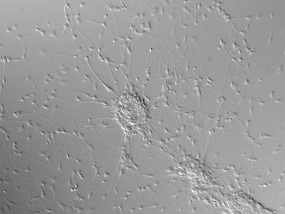
Neurons (20x) in brightfield mode suffer from low contrast.
|
Differential Interference Contrast (DIC) considerably increases the neurons’ visibility.
|
|
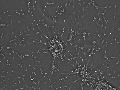
Also phase contrast adds a lot of contrast to the image.
|
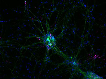
Fluorescence microscopy enables imaging of distinct proteins. Here: Nestin (green), Doublecortin (pink), nuclei (blue)
|
|
Specimens for brightfield microscopy commonly have to be stained e.g. with Hämatoxylin-Eosin (H&E). This is a part on an aorta (20x).
|
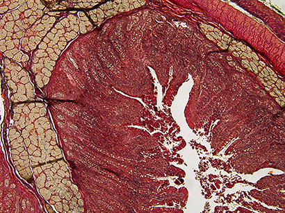
Cross-section of a duodenum in brightfield illumination (20x). LED illumination provides a constant color temperature .
|
|
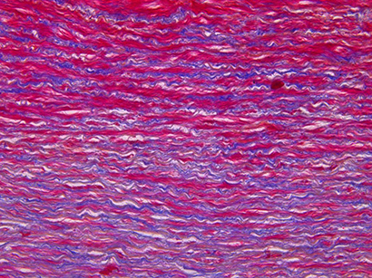
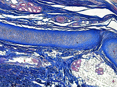
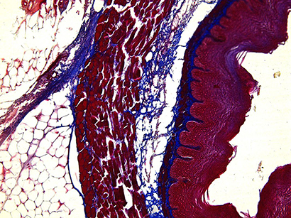
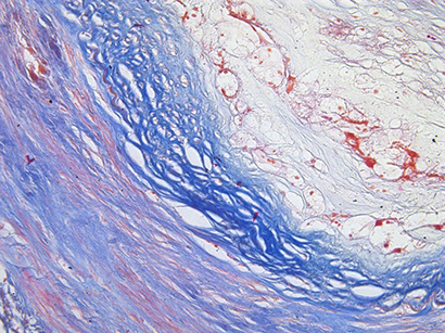
Azan staining is often used to mark connective tissue e.g. of the ear. Brightfield (5x)
|
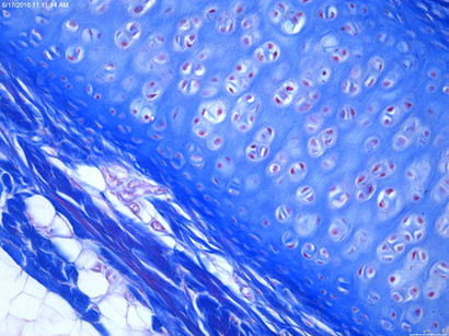
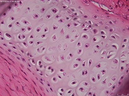
Azan staining of the ear with higher magnification (20x). Brightfield
|
|
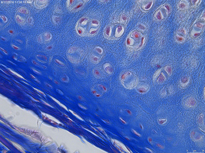
Azan staining of the ear with higher magnification (40x). Brightfield
|
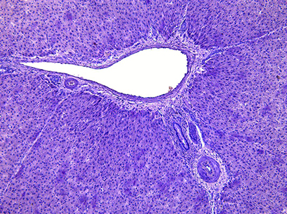
Liver tissue. Brightfield image (20x)
|
|
Oesophagus in brightfield illumination (20x)
|
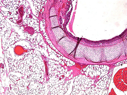
Brightfield image of a trachea (5x) stained with the well-established histological dye Hämatoxylin-Eosin (H&E)
|
|
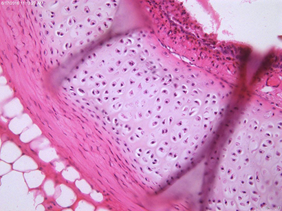
Trachea in higher magnification (20x) H&E staining, brightfield.
|
Trachea in higher magnification (40x) H&E staining, brightfield
|
|
A vessel in brightfield illumination (20x)
|