What is the right research microscope for me? This article gives you a brief overview of the major features you should have in mind when choosing an optical research microscope.
An optical microscope is often one of the central devices in a life-science research lab. It can be used for various applications which shed light on many scientific questions. Thereby the configuration and features of the microscope are crucial for its application coverage, ranging from brightfield through fluorescence microscopy to live-cell imaging.
This article provides a brief overview of the relevant microscope features and wraps up the key questions one should consider when selecting a research microscope.
One of the first things to consider when selecting a research microscope is the type of specimen you want to explore. For fixed samples mounted on a thin glass slide, you can use an upright microscope. Living cells demand special characteristics of the microscope because they are kept in relatively large cell culturevessels filled with cell culture media.
Only an inverted configuration, with the objective below and the condenser above the specimen, facilitates the essential free space and the required proximity of the objective to the specimen. At the same time an inverted microscope keeps a good accessibility to the cells, e.g. to add micromanipulators.
In addition, living cells require an adequate environment to survive. Temperature and CO2concentration have to be kept at certain levels. A climate chamber with the corresponding controllers is necessary to fulfill this task.

Figure 1: Left: Glass slide to mount fixed samples e.g. histological sections. Right: Petri dish for cell culture usage.
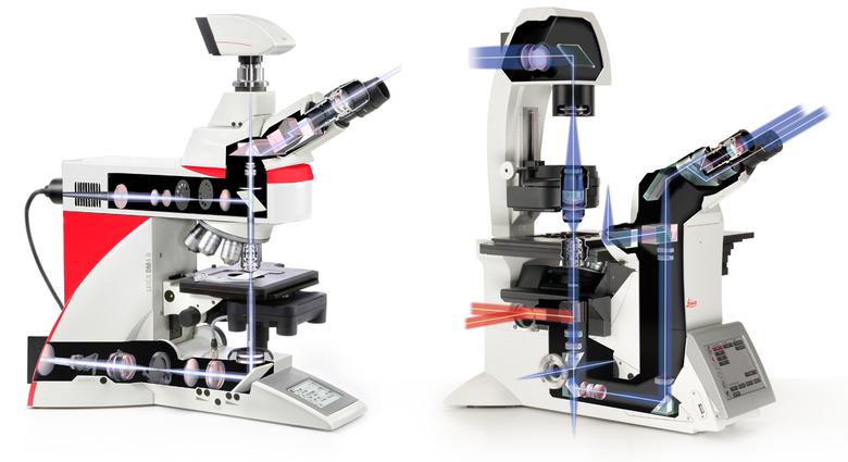
Figure 2: Left: An upright microscope features the objective above and the condenser below the specimen. Right: On an inverted microscope this set-up is turned around giving the users more space plus the required proximity of the objective to the specimen.
Microscopic specimen spread out into three dimensions: length, width, and height. Whereas some specimens, such as histological sections, are imaged only in xy-direction, there are other applications demanding acquisition also in z-dimension. To image 3D volumes e.g. of living cells, a motorized objective revolver is recommended which is able to guide your sample stepwise through the focus. The imaging software should be able to reconstruct the single images for 3D visualization.
For living cells you have to add the dimension time. In this case, for example system stability is another critical feature. Due to the fact that temperature changes influence the imaging system during acquisition, effective counter measures are essential. An automatic focus adjustment such as the Adaptive Focus Control (AFC) counteracts these thermal influences and always finds the predefined focus.
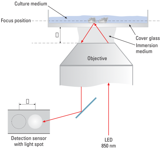
Figure 3: The Adaptive Focus Control (AFC) automatically stabilizes the microscope focus also during long-term time-lapse acquisition. A sensor detects movements of an LED light beam (850 nm) which occur if the cover glass carrying the specimen changes its position e.g. due to thermal activity.
The majority of cells – especially animal cells – investigated with a microscope don’t have enough intrinsic contrast to see fine details. Researchers use contrast methods to fix this problem. Whereas phase contrast (PH) and differential interference contrast (DIC) manipulate the light passing through the specimen to add contrast, you can also stain it with fluorescent dyes (Immunofluorescence) respectively use fluorescent proteins.
According to the contrast method the microscope needs specific equipment; e.g. phase contrast needs special objectives whereas DIC utilizes certain prisms which have to be switched into the light path. For fluorescence microscopy you need special filter cubes allowing the correct light wave lengths to access and exit the specimen.
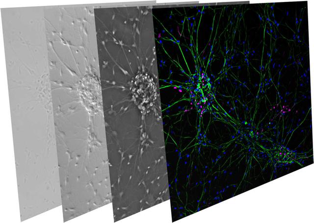
Figure 4: Series of neuronal cells acquired with different contrast methods. From left to right: Brightfield, DIC, Phase contrast, Fluorescence
The choice of the contrast method also determines the light source. Transmitted light illumination for conventional brightfield microscopy, phase contrast and DIC can be carried out with halogen or LED illumination. Fluorescence microscopy can be either performed with LED illumination or with the help of mercury, xenon, or mercury metal halide lamps.
If you want to take an image of your specimen or do live-cell imaging, you need a digital microscope camera. Especially in case of fluorescence live-cell imaging a sensitive camera is recommended to minimize the amount of excitation light which can harm the cells. In addition to the well-established CCD and EMDDC cameras, nowadays sCMOS cameras got in place due to their high quantum efficiency and acquisition speed. For more information about digital microscope cameras please read the article Introduction to Digital Camera Technology.
Furthermore, a large Field of View (FOV) helps to find interesting areas more quickly and to image more cells at the same time. Modern research microscopes feature a 19 mm FOV at the camera port which matches a 19 mm sCMOS camera chip perfectly.
Often, it is not sufficient to only take an image of your specimen, but to analyze the data you acquired. For this purpose easy-to-use imaging and analysis software helps to get quantitative data and to do solid data analysis.
During the last years, photo-manipulation of the specimen became popular. That means researchers not only watch the living cells but manipulate them with the help of light. Fluorescence Recovery after Photobleaching (FRAP) is one example which helps to untangle dynamic cellular processes. For these kinds of manipulation techniques often additional light sources are needed which have to be integrated into the microscope’s light path.
This approach is not trivial. The Leica Infinity Port is a universal solution which couples additional light sources into the microscope’s light path without disturbing image quality to do e.g. FRAP, photo-switching, ablation, or optogenetics. With the right adapter at hand, researchers can even couple their homebuilt devices.
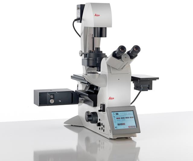
Figure 5: The Leica WF FRAP module (black box on the left) can be connected with the inverted research microscope Leica DMi8 via the Infinity Port.
One important question is how much money you can spend. Some microscope suppliers offer predefined configurations which are fit to special applications. But what if you don't need all of the preconfigured components you pay for? That's why a free configuration of components can be cheaper than buying a predefined microscope system.
Furthermore, the requirements for a microscope might change with time. In this case an upgradable system has certain advantages. With a predefined and fixed configuration you can find yourself tied to a limited amount of applications. Upgradability gives you the freedom to grow with changing demands.
Taking these points into account, a modular microscopy platform, such as the Leica DMi8, enables the researcher to start with an affordable microscope system which can be upgraded later on and grow with his demands.
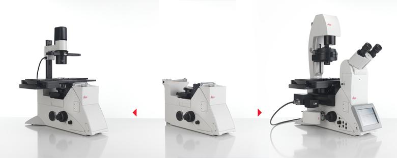
Figure 6: Thanks to its modularity, the Leica DMi8 can be configured according to the researchers needs. Moreover it can be upgraded later on, if the requirements have changed.
The range of microscope users can be very inhomogeneous. Especially at the university users can be very experienced or absolute beginners. Thus, an easy to use microscope system run by an intuitive software, such as the Leica Application Suite X (LAS X), helps to get people started quickly and to acquire data rapidly. For example a workflow oriented design, image analysis wizards, and a seamless integration of peripherals into the system simplify your work.
Besides widefield research microscopes, stereo microscopes are also often used in life-science research labs. Please find more information in the article "Factors to Consider When Selecting a Stereo Microscope".