Trace elements and their spatial distribution in rocks and minerals are of substantial interest to geoscientists: for example in igneous rocks, trace elements are valuable
tracers for magma sources and geological processes affecting the subsequent evolution of magma. Electronbeam techniques using WDS are state-of-the-art tools for
such in-situ investigations [1].
Here we present a study on major and trace element distributions in plagioclase (sodium-calcium feldspar), the commonest rock-forming mineral series. Strontium (Sr), a trace element in plagioclases, is of special interest since it can be used for diffusion chronometry or for studies of crystal-melt evolution [2], [3].
The analytical challenge in this specific case is not only related to the low concentration of the element of interest (Sr < 1000 ppm), but also to the distinct EDS peak overlap of Si Ka and Sr La.
Sample
The investigated sample is a volcanic rock from Savo in the Solomon Islands [4]. Savo is an island arc stratovolcano, with two thirds of the cone below the sea and one third exposed as the island. The sample is a trachyte (crystal rich volcanic rock with a sodium-dominated intermediate composition) from the main crater. Though the exact age of the sample is not known, much of the exposed material at Savo would have been erupted in the last 400 years.
The eruption style is that water-rich magmas ascend through the crust, crystallizing across a range of pressures and temperatures. In the shallow subsurface they presumably degas and have a final period of crystal growth, before they are “squeezed out” as crystal-rich lavas into domes which commonly collapse or explode to
produce large debris flows.
Measurement conditions
The measurements were performed on a FE-SEM equipped with both, a Bruker XFlash® ED spectrometer and a Bruker XSense® WD spectrometer using a grazing incidence parallel beam optic. The trace element Sr was determined by WDS, all other (major) elements were determined by EDS. Matrix corrections were made by
ϕ(ρZ).
Table 1 compiles the measurement conditions.
|
Measurement parameter |
Value |
|
Acceleration voltage |
15kV |
|
Probe current |
90nA |
|
EDS detector |
XFlash® 6 l 10 |
|
Outgoing count rate |
480 kcps |
|
WDS detector |
XSense® |
|
X-ray line by WDS |
Sr Kα |
|
Diffractor crystal |
PET |
|
Acquisition time |
10 h |
Results
The porphyritic volcanic rock contains abundant feldspars, varying from sodium-rich (albite, NaAlSi3O8) to relatively calcium-rich (labradorite, Na0.4Ca0.6Al1.6Si2.4O8) plagioclase compositions. Fig. 1 gives an impression of the porphyritic texture with plagioclase as dominant phenocryst phase.

Fig. 1 Element distribution map for Al, K and P in the investigated sample. High Al contents characterize the plagioclases, whereas K indicates the surrounding matrix and some biotites. Apatite is obvious due to its high Ca content. The EDS data was acquired simultaneously with WDS.
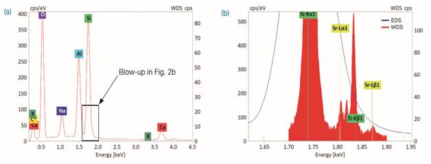
EDS spectra of such plagioclases show prominent peaks for Si, Al and O besides minor, variably high peaks for Na and Ca (Fig. 2a). Although Sr may partition in trace amounts into plagioclases, it cannot be determined from the EDS spectra.
This predicament is due to the coincidence of:
Using elaborate peak deconvolution methods, EDS is, however, able to detect Sr upon manual interaction of the skilled analyst. Nevertheless, count rates are very low and quantification of a trace element such as Sr overlapping with a major element such as Si yields inaccurate results with large errors.
In contrast, WDS is able to detect even Sr L peaks of low intensity in the X-ray spectra of plagioclase (Fig. 2b). The energy range scan obtained with WDS clearly shows the Sr La peak (1.806 keV) right inbetween Si Ka (1.740 keV) and Si Kb (1.837 keV).
The resolution of the applied QUANTAX WDS at the energy region of interest is 3.5 eV (FWHM of Si Ka) which means an improvement in resolution by a factor of 20 relative to EDS. The intensity ratio of the Sr La and Si Ka peaks is 1:170 for the present WDS spectrum in Fig. 1b. The small peak at 1.819 keV is Si Kb’, a low energy satellite line of Si Kb.
Applying a peak-background method and SrF2 as reference material for standard-based quantification of selected point analyses, Sr concentrations of up to 0.1 wt.% were determined by WDS for the most calcic plagioclases.
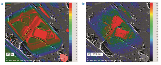
Plagioclase phenocrysts within the studied rock frequently show multiple and sometime complex zoning (Fig. 3). High contents of Ca (labradorite, Na0.4Ca0.6Al1.6Si2.4O8) are characteristic for the crystals core and may recur repeatedly throughout the zoned crystals. The plagioclases get more Na-rich (albite, NaAlSi3O8) towards their rim. Zones with high contents of Na or Ca are inevitably inverse.
X-ray spectra of a plagioclase

Fig. 2 X-ray spectra of a plagioclase in the investigated sample derived by (a) EDS and (b) combined EDS-WDS analysis. Fig. 2b shows an enlarged section of the X-ray spectrum in the region of interest between 1.6 – 1.95 keV, where the EDS spectrum is presented in light red and the WDS energy range scan in brown.
Spatial distribution of the trace element Sr within the plagioclases was determined by WDS mapping. The respective compositional maps (Fig. 3a, b) consistently show
that the highest Sr concentrations correlate well with the Ca-rich zones in the plagioclase crystals.
Discussion
The plagioclases in the present sample record crystal growth during magma evolution and ascent, and are often spectacularly zoned from calcic to sodic compositions. Disequilibrium compositional zonation of igneous minerals are commonly explained by derivation from a long-lived magma system with multiple replenishment by mafic mantle melts [1]. Whereas high contents of Ca indicate juvenile magma injected into the magma chamber and contributing to the growth of the plagioclase crystals, the crystals get more Na-rich during subsequent fractional crystallization [5].
The present results on the distribution of Sr substantiate that trace amounts of strontium can substitute for calcium in the crystal lattice of plagioclases. Moreover, the findings indicate that the juvenile magma which was involved in the formation of the present volcanic rock was fairly enriched in Sr.
Compositional maps of a plagioclase

Fig. 3 Qualitative compositional maps of a complexly zoned plagioclase presented in palette colors (map size 314 μm x 245 μm). Map (a) shows the distribution of the major element calcium derived from EDS data. Map (b) shows the distribution of the trace element strontium simultaneously acquired by WDS.
Conclusion
Electron-beam techniques allow high-resolution imaging and quantitative analysis of compositional zoning in minerals. The results clearly show that QUANTAX EDS
and QUANTAX WDS are powerful tools for the analysis of geological samples, such as plagioclases.
Installed on the same (FE-)SEM, both spectrometers (EDS and WDS) ideally complement each other. QUANTAX WDS is perfectly suited for mapping and in-situ
analyses of the trace element Sr in silicates. The WDS technique is indispensable especially under such analytical circumstances where the close proximity of X-ray lines coincides with pronounced differences in line intensities.
References
[1] Ginibre, C., Wörner, G., Kronz, A. (2007): Crystal zoning as an archive for magma evolution. Elements, 3(4), 261-266.
[2] Blundy, J. D., Wood, B. J. (1991): Crystal-chemical controls on the partitioning of Sr and Ba between plagioclase feldspar, silicate melts, and hydrothermal solutions. Geochim. Cosmochim. Acta, 55, 193-209.
[3] Zellmer, G. F., Sparks, R. S. J., Hawkesworth, C. J., Wiedenbeck, M., (2003): Magma emplacement and remobilization timescales beneath montserrat: Insights from Sr and Ba zonation in plagioclase, Phenocrysts. J. Petrol., 44, 1413-1431.
[4] Smith, D. J., Petterson, M. G., Saunders, A. D. et al. (2009): The petrogenesis of sodic island arc magma at Savo volcano, Solomon Islands. Contrib. Mineral Petrol., 158, 785.
[5] Deer, W. A., Howie, R. A., and Zussman, J. (1992): An introduction to the rock-forming minerals, 2nd Ed., Longman, London.
Acknowledgements
We thank Dr. Daniel J. Smith, University of Leicester, UK, for providing the thin section sample and detailed information on the sample location.
Author
Dr. Michael Abratis, Senior Application Scientist WDS, Bruker Nano GmbH, Berlin, Germany