|
See Beyond
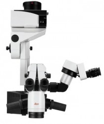
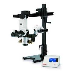
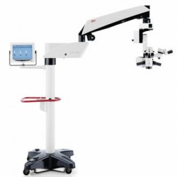
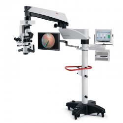
Ophthalmic Microscope Proveo 8
In the most critical moments of ophthalmic surgery, you need to be able to rely on consistent, uncompromised images, because you can’t treat what you can’t see.
The Proveo 8 ophthalmic microscope goes beyond conventional visualization. Its exclusive optical technology provides you with both constant red reflex and a rich texture view, throughout entire anterior and posterior procedures.
Proveo 8 pushes the boundaries of visualization:
• CoAx4 Illumination technology for enhanced view during all cataract surgery stages, including phacoemulsification
 Liên hệ với chúng tôi Liên hệ với chúng tôi
|
 |
See Every Detail
As a posterior segment surgeon, you need to carry out extremely precise work from the edge of a detached membrane layer to the retina, often in low light. Until now, this meant time-consuming refocusing, and limitations in image clarity and detail.
Exclusive FusionOptics technology delivers crisp, texture-rich images from the periphery to the membrane layers, enhancing your efficiency and precision.
The Technology of FusionOptics 1. Two separate beam paths2. One beam path provides 40% increased depth of field 3. The other beam path provides high resolution 4. The brain merges the two images to a single optimal spatial image |
Stable Red Reflex, Consistent Images
Focus on your cataract surgery and be confident that you’ll have an constant, uncompromised view.
You can rely on stable red reflex and optimal image contrast thanks to CoAx 4, coaxial LED illumination.
Plus, all observers – surgeon, assistant, and camera – work with the same enhanced view during the entire surgical procedure.
|
 |
 |
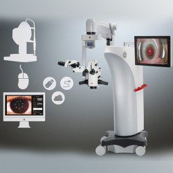
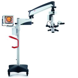
Beyond Tomorrow
Stay ahead, stay flexible. Proveo 8 delivers state-of-the-art solutions today and the ability to integrate new technologies tomorrow. The streamlined yet modular design of the Proveo 8 microscope offers the flexibility to integrate different imaging and documentation solutions.
• Let IOL guidance software support your pre-operative planning and accurate IOL placement • Visualize subsurface ocular microstructures in anterior and posterior surgery with OCT • Operate "heads up" looking at a 3D monitor and benefit from enhanced workflow, comfort and teaching |
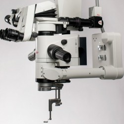
Efficiency Redefined
The best workflow is the one that just flows without interruption, because the equipment is ready for the next step and reliable in every single moment of the surgery.
• Experience efficiency by pre-programming settings according to procedure and surgery phase, then altering via footswitch – without interrupting the workflow • Work with confidence by confirming your settings at one glance to the surgeon information panel • Maneuver with ease thanks to the 21% longer reach and 33% smaller footprint*
*Compared to similar products (November 2015) |
|
Hiện tại chưa có ý kiến đánh giá nào về sản phẩm. Hãy là người đầu tiên chia sẻ cảm nhận của bạn.


 info@redstarvietnam.com