Easy to Use: Compact and portable, with incredibly simple operation.
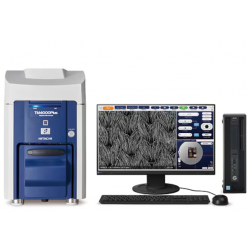
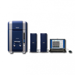
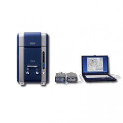
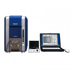
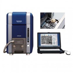
The space saving and lightweight design of TM3030 means it can be conveniently installed on a table*. No cooling water is needed, so installation is quick and easy and requires only a standard 100-240 V AC power supply.
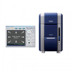
Imaging with the TM3030 couldn’t be simpler. Pressing the “Start” button automatically turns on the beam, adjusts focus, brightness and contrast, as well as displays the image at an easy-to-view starting magnification of x100.
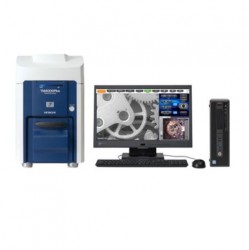
No sample Preparation; Versatility is assured - with a wide magnifi cation range and multiple
operating conditions.
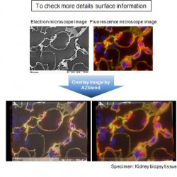
When a non-conductive sample is observed with a high-vacuum SEM,electrons accumulate on the specimen surface causing a charge-up phenomenon. Charging prevents imaging. In order to resolve the charge, the sample is usually coated with a thin layer of metal prior to observation. This process is not only time consuming, but also interferes with optical imaging of surface details as well as EDX analysis. The TM3030 overcomes this problem with “charge-up reduction mode.” This mode uses low-vacuum functionality to dissipate the charge.
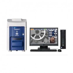
Unparalleled Image Quality; Image quality being sharpened and enhanced.
The 5kV accelerating voltage allows for observation of surface details, it not only offers traditional topographic imaging but also compositional imaging information. The 5 kV observation condition is further enhanced throughout high magnifications by improving the electron optics.
These functions will be utilized to enhance image quality at any observation condition modes and will be very effective for higher magnification specimens.
|
Items |
Description |
|
Magnification |
15 to 60,000×(digital zoom: 2×, 4×) |
|
Observation condition |
5kV/15kV/EDX |
|
Observation mode |
Standard mode |
|
Image mode |
COMPO/Shadow 1/Shadow 2/TOPO |
|
Sample stage traverse |
X: ±17.5 mm, Y: ±17.5 mm |
|
Maximum sample size |
70 mm in diameter |
|
Maximum sample height |
50 mm |
|
Electron gun |
Pre-centered cartridge filament |
|
Signal detection system |
High-sensitive semiconductor |
|
Auto image adjustment function |
Auto start, Auto focus, Auto brightness/contrast |
|
Frame memory |
640 × 480 pixels, 1,280 × 960 pixels |
|
Image data memory |
HDD of PC and other removal media |
|
Image format |
BMP, TIFF, JPEG |
|
Data display |
Micron marker, micron value, date and time, image number and comments, Image mode, Observation condition, D* (Distance), Observation mode |
|
Evacuation system (vacuum pump) |
Turbomolecular pump: 30 L /s × 1 unit, |
|
Operation help functions |
Raster rotation, Magnification preset (two steps) |
|
Safety device |
Over-current protection function, built-in ELCB |
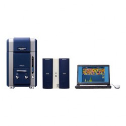
Optional accessories
Energy Dispersive X-ray Spectrometer TM3030/TM3000
|
|
Quantax70
(Manufactured for Hitachi High-Technologies corporation by Bruker Nano GmbH)
|
|
|
SwiftED3000
(Manufactured for Hitachi High-Technologies Corporation by Oxford Instruments Analytical Ltd.)
|
Optional Accessory for Tabletop Microscope TM3030/TM3000
|
|
|
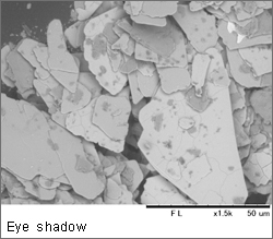
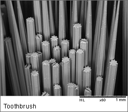
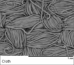
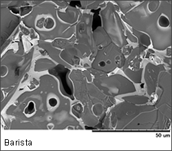
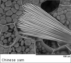
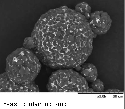
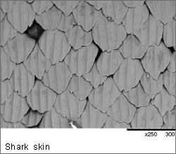
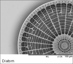
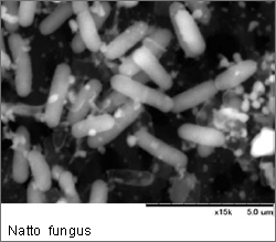
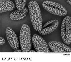
Image Gallery
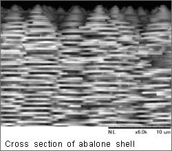
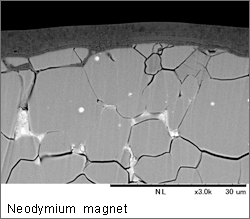
Materials
|
|
|
|
|
|
|
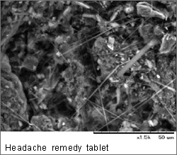
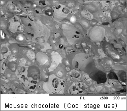
Food and Medicine
|
|
|
|
|
|
|
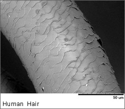
Organism
|
|
|
|
|
|
|
|
|
|
|
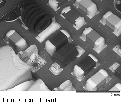
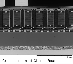
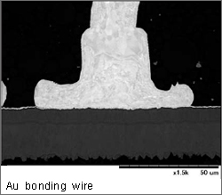
Electronic Materials
|
|
|
|
|
|
|
Hiện tại chưa có ý kiến đánh giá nào về sản phẩm. Hãy là người đầu tiên chia sẻ cảm nhận của bạn.