True MultiColor Laser TIRF Leica AM TIRF MC
|
||
|
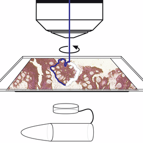
Specimen collection by gravity – contact-free and contamination-free. Laser Microdissection (LMD) makes it possible to distinguish between relevant and non-relevant cells or tissues. It enables the researcher to obtain homogeneous, ultra-pure samples from heterogeneous starting material.
Researcher can selectively and routinely analyze regions of interest down to single cells from all kinds of tissues, even living cells from cell culture, to obtain results that are relevant, reproducible, and specific.
Leica Microsystems offers two systems for LMD:
Learn more about Laser Microdissection applications on the knowledge portal on microscopy, Leica Science Lab.
 Liên hệ với chúng tôi Liên hệ với chúng tôi
|
|
|
Your Advantages

Laser Specifications Leica LMD7000Max. pulse energy: 120 µJ |

Laser Specifications Leica LMD6500Max. pulse energy: 70 µJ |

Specimen Collection by GravityLaser microdissection uses a laser to isolate specific microscopic regions from tissue samples. The way in which specimens of interest are transferred and collected influences the specificity and quality of the end result. Only Leica Microsystems uses simple gravity for specimen collection. |

Laser Movement via OpticsOnly Leica Microsystems uses high-precision optics to steer the laser beam using prisms along the desired cut line on the tissue. Benefits are highest possible precision and speed, direct real-time laser cutting with “Move and Cut” and the best view for convenient movie documentation. |

Leading-edge Laser TechnologyThe Leica LMD7000 is the first laser microdissection system that integrates high energy per pulse, and high and adjustable repetition rates within one single system. Benefits are full control of the repetition to adapt the laser to a specific sample, high-energy per pulse for thick and hard specimens, high repetition rates for narrow cuttings and high speed and control of all laser parameters including laser aperture to achieve an optimal cutting line. |

Standard devices for dissectate collectionLaser microdissection from normal glass slides is possible with the „Draw + Scan“ or directly live with the „Move + Cut“ mode. This kind of application is called laser ablation or dot scan dissection. Laser microdissected samples can be easily collected by gravity to all common molecular biology reaction devices caps like 0.2 or 0.5 ml tubes. This method is efficient and time-saving – no additional transfer steps are needed. Collection devices can be dry or with reaction buffer. No expensive special consumables are needed for collection. |

Live Cell Cutting (LCC)Cell culture plays an important role in Science. Leica Microsystems offers dedicated equipment for laser microdissection of living cells in culture in order to recultivate, clone or analyze single cells or cell clusters. If appreciated a climate chamber can be attached to the laser microdissection system.
For laser microdissection cells can grow on petri dishes with PEN membrane, stackable membrane rings or multi-well ibidi slides; culture media must be removed (a slight film may remain on the cells) prior dissection. Dissectate of living cell culture can be collected into petri dishes with or without PEN membrane, ibidi slides, stackable membrane rings or 8-stripe tubes for re-cultivation or into collection devices as PCR tube caps or LOC for analysis |

Membrane Slides & DIRECTOR® SlidesLeica provides a wide range of different membrane slides offering the best solution for your laser microdissection application. For glass slides PEN (polyethylene naphthalate) and PPS (polyphenylene sulfide) membranes are available, suitable for almost all applications (Usable area for LMD 19 x 41 mm). A special big glass slide with PEN membrane offers an usable area of 39 x 45 mm for laser microdissection for big tissue samples.
Beside the glass slides with PEN or PPS membrane (MembraneSlides), membranes on steel frames (FrameSlides) allow the usage of a special LMD 150x dry objective. Available for steel frames are PEN and PPS membrane, suitable for almost all applications, PET (polyethylene terephthalate) membrane, recommended for MALDI-TOF downstream applications, POL (polyester) membrane recommended for chromosome spreads a in combination with the 150x objective and a special FLUO membrane without auto-fluorescence and best suitable for DIC optics.
Tissue ablation with the LMD “Draw & Scan” mode enables you to collect your sample direct from a special coated, energy absorbing glass slides (DIRECTOR®) as well as from usual glass slides. In combination with the LMD “Draw & Cut” mode, the DIRECTOR® slides allow a fast and accurate sample collection with low laser power. |
Hiện tại chưa có ý kiến đánh giá nào về sản phẩm. Hãy là người đầu tiên chia sẻ cảm nhận của bạn.