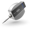


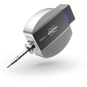
Đầu dò QUANTAX Micro-XRF
Trace Element Sensitivity with Minimal Sample Preparation
Liên hệ
10ppm
detection limit
Trace element analysis is possible thanks to a low spectral background
Minimal sample prep
Easy sample preparation
No carbon coating required and no charging effects
40µm
layer thickness
Thin films starting from 1 nm up to multiple layer structures of 40 µm can be analyzed
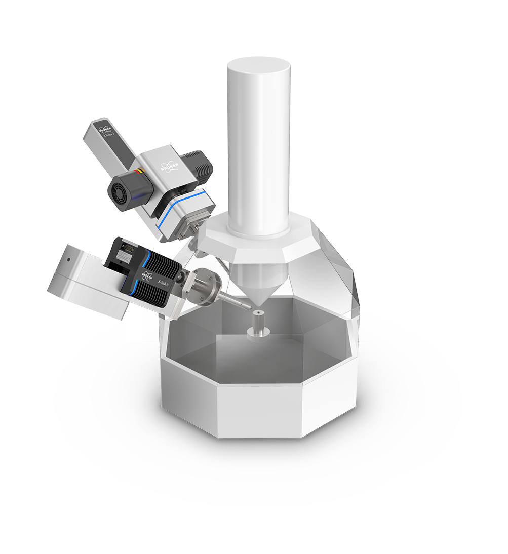
Expand your SEM Capabilities – Augment SEM EDS with micro-XRF Analysis
Micro-X-ray Fluorescence (micro- XRF) spectroscopy is a non-destructive analytical technique that can be used alongside conventional Energy Dispersive Spectroscopy (EDS) on a Scanning Electron Microscope (SEM).
Micro-XRF on SEM, also known as SEM XRF, delivers the SEM with a range of new capabilities such as the ability to measure and map trace elements, the analysis of layered samples, and more.
Trace Element Detection
X-ray analysis is more sensitive towards trace elements, allowing their detection at concentrations down to as low as 10 ppm for certain elements, an extended X-ray spectral range (up to 40 keV), as well as information from a greater depth within the sample.
Elemental Mapping with Small Spot Sizes
QUANTAX micro-XRF is equipped with XTrace 2, our latest X-ray source, including a micro-focusing X-ray optic that yields small spot sizes of down to 10 µm at a high-intensity throughput.
Multilayer Sample Analysis
The larger depth of X-ray excitation allows for the characterization of multilayer samples from 1 nm up to 40 µm, which is not possible via electron beam excitation.
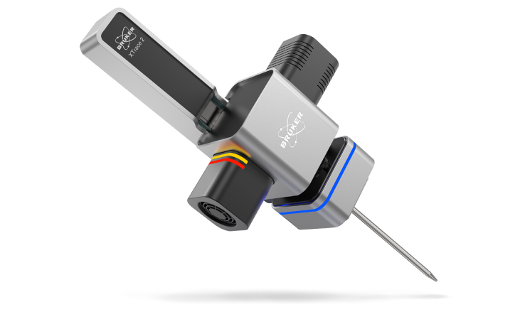
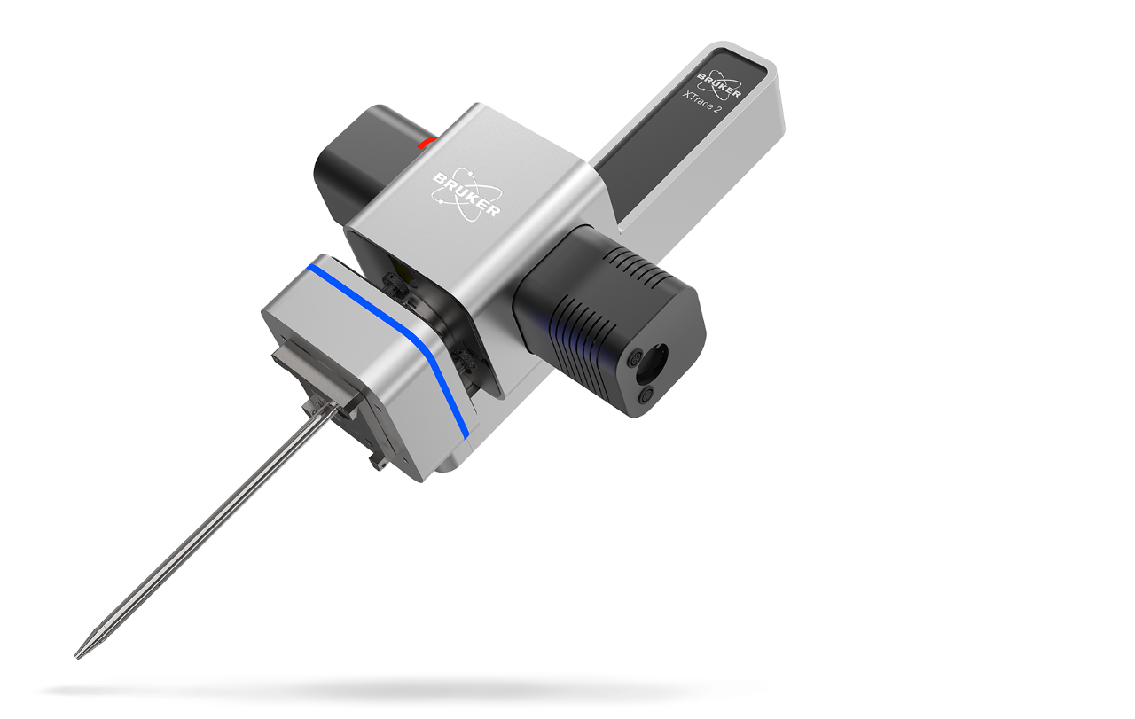
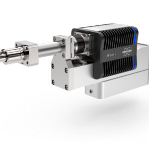
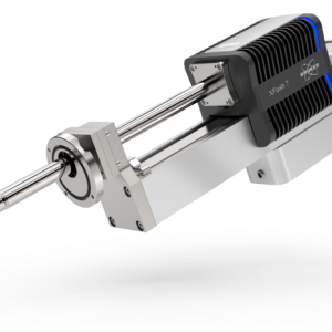
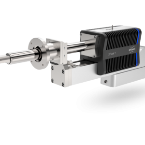
XTrace 2 – Advanced X-Ray Source for micro-XRF on SEM
XTrace 2 is the next-generation X-ray source in the QUANTAX micro-XRF system for micro-XRF on SEM (SEM XRF). This new and innovative X-ray source enables fast micro-XRF spectral acquisition with high-resolution data.
Advanced features, such as a FlexiSpot mode, an Aperture Management System, and motorized filter selection facilitate the collection of rich data from even challenging samples.
- Collect accurate elemental data quickly and efficiently with high-energy X-rays of 50 kV and beam currents of 1000 µA for a high count rate.
- Detection of trace elements at low ppm levels with automatic switching between 6 primary filters for enhanced background reduction.
- Scan topographic samples in high resolution using an Aperture Management System (AMS) that keeps the image in focus across a variable working distance.
- Analyze inhomogeneous and/or irregular samples using FlexiSpot mode, allowing spectral measurements from a small to a larger spot size.
- Retract the X-ray optic when not needed by using a motorized linear stage with automatic source retraction (measuring and parking position).
- Return to areas of interest on your sample by saving measurement positions for correlating micro-XRF / e-beam analysis.
- Maximize X-ray tube lifetime with by automatic X-ray tube warm-up procedure.
- Control the analysis, select filters, and move the linear stage using the intuitive ESPRIT software.
Aperture Management System (AMS) for the Analysis of Topographic Samples in the SEM
XTrace 2 is equipped with the patent protected AMS to retain X-ray spot resolution even when scanning samples with a complex topography in the scanning electron microscope.
The AMS in XTrace 2 keeps the optic in focus at varying working distances by increasing the depth of field. This means that any reduction in resolution due to deviations in the working distance will be minimized, facilitating the high-resolution elemental mapping of highly topographical samples and their 3D features.
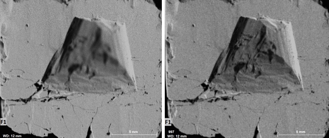
The AMS feature of XTrace 2 allows samples with a high topography to be analysed via SEM. On the left hand side is a Pyrite (FeS2) sample imaged with no AMS, on the right hand side is the same sample imaged with the AMS set to 500 µm.
FlexiSpot – Variable Spot Sizes
FlexiSpot allows users to not only take measurements from small spot sizes (10 μm, 35 μm) but also a range of larger spot sizes (50 – 500 μm). FlexiSpot works by retracting the X-ray source, allowing the X-ray optic to be defocused out of the nominal optic working distance. Users can select between spot sizes using the automated process in our ESPRIT software.
The ability to measure larger spot sizes allows for a more precise quantification of non-homogeneous and irregular shaped samples, as well as samples with uneven surfaces, such as powders. A large spot area provides more statistically accurate data on a sample’s composition in just one measurement.
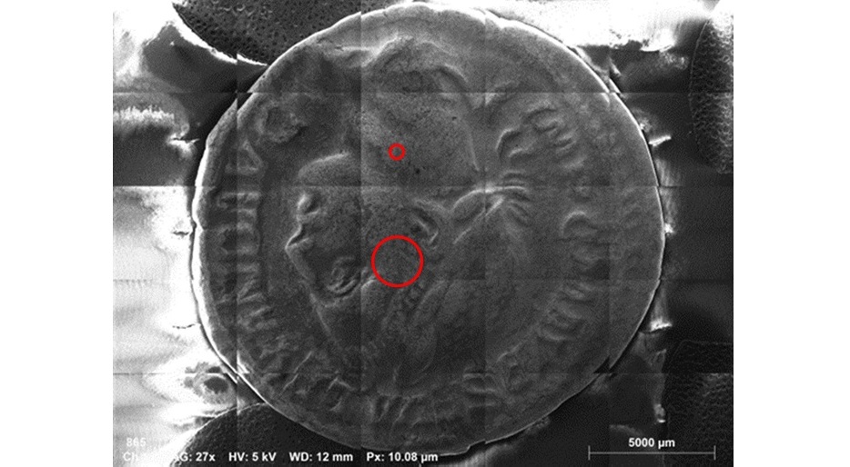
Small (35 µm) and large (200 µm) X-ray spots on a sample – FlexiSpot allows for the analysis of specific features using a small spot size and for the overall analysis of a sample using a large spot size.
Easy Switching between up to 6 Primary Filters for Further Background Reduction
The new XTrace 2 comes with 6 primary filters allows the user to adapt the background over the whole energy range up to 40 keV for further improved sensitivity.
Additional filtering allows for the reduction of the background for specific measurements at energies up to 40 keV. Primary filters can be selected via our ESPRIT software.

Auto Source Insertion & Retraction Mode
The polycapillary optic of the X-ray source can be automatically inserted and retracted using XTrace 2’s motorized linear stage.
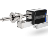
Sản phẩm tương tự
Liên hệ
Liên hệ

Liên hệ
Liên hệ

Liên hệ
Liên hệ
Liên hệ