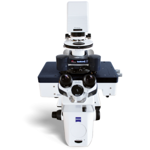
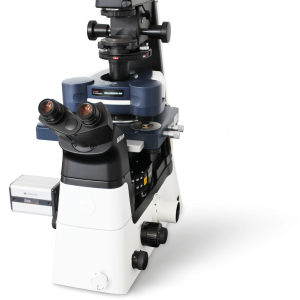
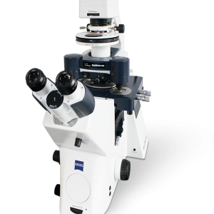
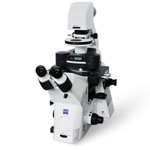
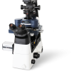
ForceRobot 400
Liên hệ
ForceRobot 400
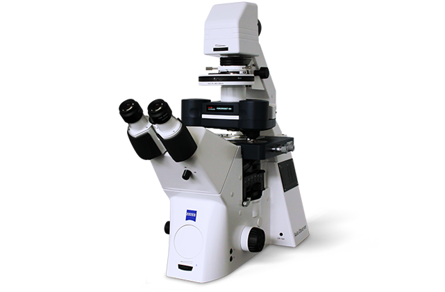
Forces play a crucial role in molecular mechanisms, such as recognition, response, and signaling. The ForceRobot® 400 BioAFM incorporates a number of unique force spectroscopy innovations to measure forces at the single-molecule level. It enables the quantification of the mechanical strength of individual molecular bonds and characterization of the force-dependent properties of molecular interactions and biomolecular complexes.
Understanding the Mechanics of Single Molecules
ForceRobot 400 is uniquely capable of providing crucial insights into functional biological mechanisms, opening new possibilities in such research fields as biophysics, biochemistry, developmental biology, and the development of novel therapeutic molecules.
ForceRobot 400 delivers:
- Outstanding performance: 250,000 force curves per day
- Fully automated single-molecule force spectroscopy (SMFS) capabilities
- Statistically relevant, standardized datasets required in biomedical and preclinical research
- Advanced force curve designs and extensive fitting routines for flexible experiments
Innovative multiparametric nanomechanical mapping
ForceRobot 400 enables the label-free, nanomechanical measurement of individual molecules under near-physiological conditions. Correlative, multiparametric datasets of unprecedented quality can be collected and a comprehensive range of biophysical parameters derived automatically from each dataset.

Next-Generation Force Measurement Capabilities
Advanced Automated Force Spectroscopy
ForceRobot 400 is the ideal tool for SMFS, providing crucial insights into biophysical mechanisms at the molecular level. Large-scaled z-motors and an enhanced motorized stage functionality enable the investigation of samples ranging in size from single molecules to larger biological complexes and regions of interest (ROI) that are far apart or in different sample compartments. The latest optional NestedScanner feature has a fast z-scanner with a capacitive sensor. It generates reproducible force curves even at high speeds, significantly extending the frequency range for microrheological measurements.
The new SmartMapping feature allows the flexible selection of multiple, userdefined 2D force maps. Using optical tiling, multiple ROIs can be selected in advance and examined automatically, facilitating the systematic study of large sample areas.
Setting New Standards in SMFS Automation
ForceRobot 400 delivers advanced automation and analysis capabilities at unprecedented data acquisition rates. Automated alignment and calibration features, in combination with proprietary software tools, ensure autonomous operation and rapid results.
Ultimate Flexibility
The new ForceRobot 400 user interface, with intuitive user guidance, enables the fast and simple definition of operating parameters. Pre-defined experiment setups, such as force-clamp, force-ramp, or microrheology, along with easy-to-use scripting tools allow user-defined experiment designs. Long-term, selfregulating experiment series can be run thanks to the automated adjustment of system parameters.

Groundbreaking Capabilities:
- Over 10,000 force curves per hour
- Automated alignment of detection system and adaptation to environmental conditions
- New automated self-adjusting mapping mode for probing rough surfaces
- Characterization of viscoelastic properties with a modulation frequency of at least 5 kHz (requires optional z-scanner)
- Extensive range of environmental control accessories (humidity, temperature, ionic strength, buffer exchange, etc.)
Seamless Integration with Optical Microscopes
ForceRobot 400 can be seamlessly integrated with advanced optics and super-resolution techniques to deliver correlated nanomechanical data sets and the comprehensive characterization of an extensive range of biological samples.


