


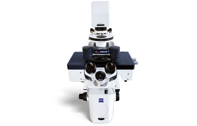
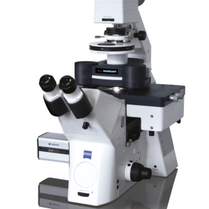
NanoWizard® 4 XP BioScience
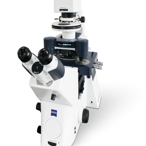
The NanoWizard 4 XP BioScience atomic force microscope combines atomic resolution and fast scanning with rates of up to 150 lines/sec and a large scan range of 100µm in one system. It is designed to provide highest mechanical and thermal stability on inverted optical microscopes during long term experiments on samples ranging from single molecules to living cells and tissues.
High-resolution imaging with extreme performance
The NanoWizard 4 XP BioScience comes with:
- PeakForce Tapping® for easy imaging
- Fast Scanning option with up to 150 lines/sec
- NestedScanner Technology for high-speed imaging of surface structures up to 16.5µm with outstanding resolution and stability
- New tiling functionality for automated mapping of large sample areas
- V7 Software with revolutionary new workflow-based user interface
- DirectOverlay™ 2 software for perfect integration and data correlation with advanced fluorescence microscopy platforms
- Vortis™ 2 controller for high-speed signal processing and lowest noise levels

Tailored DNA origami frames imaged in TAE 10mM MgCl2 buffer on mica. Sample courtesy of R. Willaert, VUB, Brussels (BE). Scan field: 125nm · Height range: 4.4nm · Scan speed: 150 lines/sec
Automated mapping of large sample areas with new tiling functionality

Living Vero cells in cell culture medium at 37°C.
The HybridStage™ or Motorized Precision Stage transforms experiments by enabling direct access to a large sample area, with automated, motorized movement to selected positions, grids and mapping regions.
Begin with the DirectOverlay 2 optical calibration, and then select a region for optical tiling up to millimeters in size.
Precise motor movements automatically bring the whole sample into view, making it easy to select regions and features for further investigation. A single click navigates from point to point or MultiScan experiments automate a sequence of measurements at selected points.
The images show living Vero cells in cell culture medium at 37°C in the PetriDishHeater™ . [2] – [5] Optical tiling with 5×6 phase contrast images covering a 630µm×450µm region. – Zoom into region scanned with AFM showing 100μm×100μm scan (height range 5μm) and inset 15μm×15μm (height range 2μm) scan topography images using PeakForce Tapping. The feedback correction signal images highlight the surface membrane features, particularly in the zoomed image. Microvilli dominate the center of the cell, with membrane ruffles at the cell boundary.
Perfect optical integration provides true correlative microscopy
The NanoWizard 4 XP, with its unique tip-scanning technology and fast imaging capabilities, is ideal for taking advantage of the synergy between AFM and super-resolution microscopy.
The NanoWizard 4 XP is compatible with a wide range of platforms, such as those from Zeiss (PALM/STORM, SIM), Leica (STED), PicoQuant (STED), Nikon (SIM, STORM) and Abberior (STED).
The 980nm laser option for the AFM head allows the simultaneous use of optical microscopes and focus stabilization systems, critical for long term experiments, and avoids conflicts with fluorescence or spectroscopy measurements.

STED and AFM experiment of living A549 cells imaged at 37°C in medium. [1] STED image of microtubules labelled with silicon rhodamine overlayed with AFM topography. [2] AFM QI topography image at 240pN imaging force (height range 3.5μm). [3] Corresponding QI Young‘s modulus image (z range 100kPa).

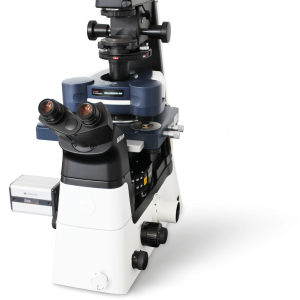
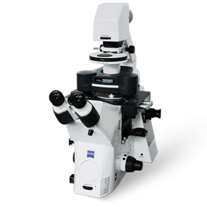
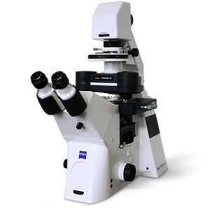
The images show NanoWizard 4 XP setups on Zeiss LSM 880 confocal microscope with Airyscan [1] , with Upright Fluorescence Microscope (UFM) Kit for tissues or other large samples such as organs [2] , with BioMAT Workstation for high NA optics and Zeiss Axio Imager [3] , with TopviewOptics [4] and on on Olympus with PicoQuant MicroTime 200 STED [5].
Outstanding quantitative data from molecules, cells and tissues
QI™ Advanced, based on real force curves, offers both astounding speed and resolution for applications ranging from single molecules to living cells. The quantitative data allows precise and fast analysis of mechanical or biochemical interactions, e.g., localization of binding sites, directly overlaid with fluorescent labelling and topography with Molecular Recognition Imaging. Advanced batch processing options include multiple models for modulus fitting and can reveal surface topography at zero force with Contact Point Imaging (CPI).
The image shows single cell force spectroscopy measurements using the CellHesion module with an increased z range of 100 µm, showing the detachment force curves of a single A549 cell from fibronectin (FN) and from bovine serum albumin (BSA) coated culture dishes. Note, that the detachment of the cell from fibronectin results in a very large pulling range of 77 µm.

Single cell force spectroscopy measurements using the CellHesion module.

[1]-[2] Stiffness mapping of non-cancerous human cervix tissue with a HybridStage. The inset in the fluorescently-labelled (Hoechst) slab was used for mapping of a 5×4 quadrant area of 1000μm×800μm with overlayed composite Young‘s modulus map shown in the middle panel. [3] The representative topography channel from an individual 200μm×200μm channel is given in the right panel. Sample courtesy of Dr. T. Fuhs and Prof. J.A. Käs, University of Leipzig, Germany.

Operating Modes
Standard Operating Modes
Imaging modes
- Now with PeakForce Tapping
- Contact mode with lateral force microscopy (LFM)
- Tapping Mode™ with PhaseImaging™
Force measurements
- Static and dynamic spectroscopy
- Advanced force mapping
Optional Modes
- Fast Scanning option with up to 150 lines/sec
- Fast QI Advanced mode for quantitative data, perfect for soft samples
- Mechanical properties such as adhesion, elasticity, stiffness, deformation
- Conductivity and charge distribution mapping
- Contact Point Imaging (CPI) with zero force
- Molecular recognition imaging for binding site mapping
- Advanced AC modes such as FM and PM with Q-control & Active
Gain Control
- Higher harmonics imaging
- Kelvin Probe Microscopy and SCM MFM and EFM (see also QI mode)
- Conductive AFM (see also QI mode)
- STM
- Electrical spectroscopy modes
- Piezoresponse Microscopy for high voltages
- Electrochemistry with temperature control and optical microscopy
- NanoLithography
- NanoManipulation
- Nanoindentation
- Scanning Thermal AFM
- FluidFM® solution from Cytosurge
- ExperimentPlanner for designing a specific measurement workflow
- RampDesigner™ for custom designed clamp and ramp experiments
- ExperimentControl feature for remote experiment control
- DirectOverlay 2 for combined AFM and optical microscopy
- Additional XY or Z sample movement stages available with CellHesion®, TAO™ and HybridStage™ module
Related products
Liên hệ
Liên hệ
Liên hệ
Liên hệ
Liên hệ
Liên hệ
Liên hệ