Color Fundus Guides Your Way
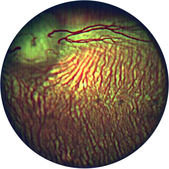
Quickly and precisely image your target region of interest with Color Fundus guidance support. The integrated EnFace™ Color Fundus camera displays bright, full-color images of the retina and cornea while the OCT scan head captures the full 3D depth of the tissue.
- Save time: Fast alignment allows you to increase your experiment throughput on small and large animals
- Explore deeper: Rapidly identify surface pathologies and quickly establish protocols to probe deeper into the targeted tissues
- See richer detail: Get high spatial resolution for maximum visualization
- Efficiently screen: The EnFace™ camera helps you to screen quickly animal eyes
- Publish in high quality: Brilliant, detailed fundus images for featuring your research
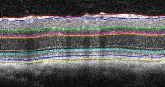
Full Volumes in One Go
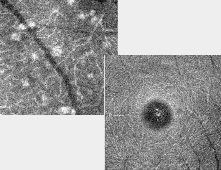
Uncover the secrets of the eye by getting the full picture with Envisu Image Guided SDOCT systems. Quickly capture the entire 3D tissue volume in exquisite detail within 3 seconds. Explore retinal layers in rich, high-resolution detail, enabling you to detect markers that indicate the earliest signs of disease. Our High Density (HD) scan management system provides you with fully customizable data scans, maximum image density, and unparalleled resolution voxels of the retina.
- Analyze HD data scans in 1000 x 1000 x 1000 with maximum image density
- Segment the layers of retina using the full volume, Diver 8-layer mouse segmentation software for early detection of pathology
- Fully registered, depth-selective fundus images are projected from OCT data, later allowing you to conduct post-imaging explorations on full volume data sets
Power Up Your Analysis
Analyze deep into your data with the fast and easy-to-use Diver Analysis Software, the only full-volume, automated retinal thickness analysis software for small animal models.
- Quickly and accurately identify all retinal layer boundaries in 3D. Diver is designed for global or regional analysis of the mouse retina.
- Conduct longitudinal measurements in the region of interest and compare changes in retinal layers.
- Harness the powerful automated 8-layer mouse retina segmentation and analysis tool for faster results. Diver’s template-based manual marking option provides further customized segmentation capability.
- Populate a spreadsheet for analytics and generate a report with statistical results and layer-wise thickness and heat maps.
From Small to Large Animal
Image any species you want with just one flexible OCT system: The Envisu Image Guidance systems provide you flexibility by providing customized lenses to fit your needs and animal model.
Get flexibility for multi-species translational research with precision posterior and anterior imaging bores for targeting small to large animals.

Our EnFace Image Guided OCTs come with a full set of species specific imaging lenses:
- Mouse retina lens with 50 degree field of view and 2.5 µm lateral resolution
- Rat retina lens with 40 degree field of view and 4 µm lateral resolution
- Rabbit retina lens with 70 degree field of view and 9 µm lateral resolution
- Anterior Imaging: Select from 10 mm and 20 mm field of view anterior imaging lenses


 nhãn khoa Envisu C-Class")