|
Leica Application Suite X
Leica Application Suite X (LAS X) is the easy to use software platform for advanced life science research on Leica Microsystems confocal, widefield and super-resolution systems.
Contact Our Software Experts
|
|
|
The Power of LAS X
Intuitive, Innovative, Indispensable.
The Leica Application Suite X (LAS X) combines the most powerful features available today in microscope software into one package, focusing on usability in every aspect of the interface, functionality and workflow.
The workflow oriented design and the possibility to recall user defined system settings makes LAS X a very intuitive imaging platform customized to your needs. Combining innovative software and hardware options like the Mobile Connection module, the LightGate and Lambda Square Mapping on White Light Laser Systems or the water immersion micro dispenser makes LAS X an indispensable solution for confocal, widefield and super-resolution microscope systems.
|
|
Easy and Versatile Wizards for 3D and 2D Analysis of Multi-Dimensional Data Sets
Obtain reproducible analysis results rapidly and easily with the 3D and 2D image analysis wizards. Go step by step through the guided workflow: from applying filters, thresholding, and binary image processing to measurements and classification. Use a binary reference mask in multi-channel image analysis, for example, to count the number of spots in each nucleus. In addition, you can perform 2D tracking experiments or call ImageJ macros from within the 2D analysis wizard.
Save the analysis results with the experiment or create a report including histogram and binary masks and export it to Excel for further analysis.
For repetitive tasks, user-defined protocols can be saved and applied to new data sets. To manage large amounts of data, several data sets can be added to a batch list and processed at once. Results from different images can also be accumulated, providing statistical information.
Your Advantages
|
|
|
|
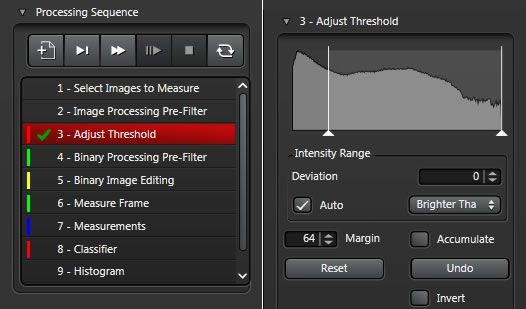
Guided Workflow
Create reproducible results step-by-step using the Analysis workflow. At each step a set of analysis tools are available. The viewer provides immediate feedback on the applied settings. Additionally, tools such as auto threshold, split objects, or edge removal can be used for automated analysis of multiple samples. When needed, you can set pauses during an automated analysis to allow you to make fine adjustments, for example for binary image editing.
|
|
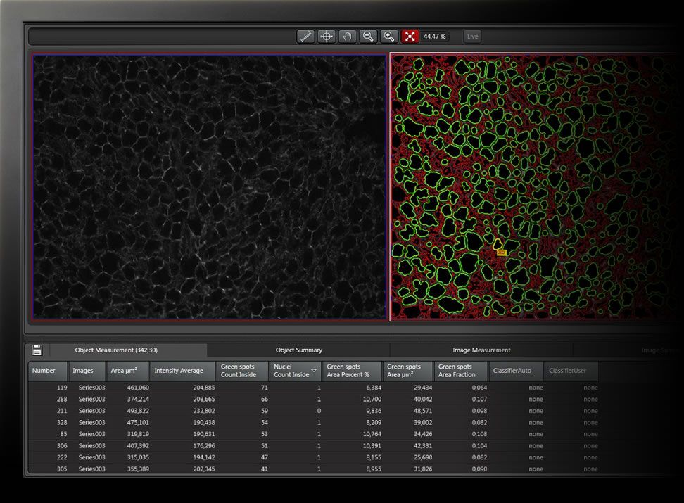
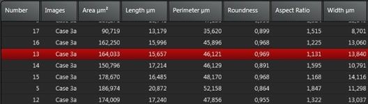
Interactive Measurement Table
Get to the data you need with the flexible and interactive measurement table. Select objects in the image viewer and the corresponding measurement is highlighted in the results table, or vice versa. Choose from a large number of measurement parameters and rearrange or sort the columns as you like. Export measurements and statistics to Excel or save them with the experiment. Reproduce measurements easily with user defined protocols.
|
|
|
|
Multi Channel Analysis made easy
With multi-channel analysis, even complicated fluorescent experiments can be evaluated with ease. Create separate analysis channels for each fluorescent marker and assign an individual workflow to each. Then simply run an analysis of all aspects of your sample at the same time.
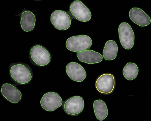
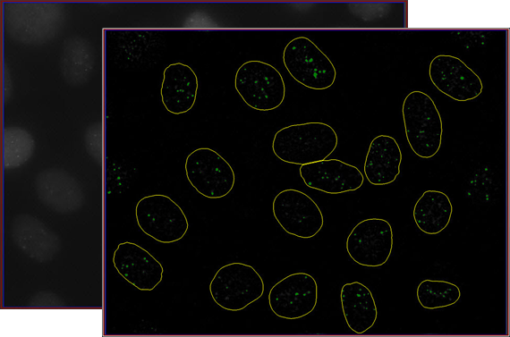
Combine analyses between separate channels to get object specific data. For example, apply a binary reference mask to count the number of spots in each nucleus.
Alternatively, apply multiple analysis channels to the same image. On a stained color image you can count the healthy cells in one analysis channel and create a second analysis channel to count the abnormal cells, for example. |
|
|
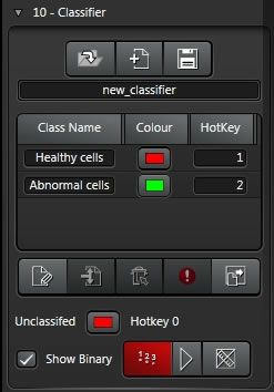
Automatic Classifier
Get unbiased and reproducible classification results even in situations where thresholding or filtering is not suitable. Use the intelligent classifier to group your results and display them in a histogram. The classifier can be trained with a training data set, then saved and applied to new data sets. The new objects will be divided into your predefined classes with a support vector machine.
|
|
Follow your particles
Perform 2D tracking within the 2D Analysis wizard. Display particle trajectories, follow the objects along their trajectories over time and export the tracking results to excel.
|
|
|
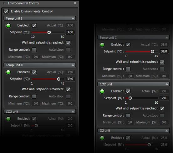
Make Temperature, CO2 and O2 Conditions Part of your Experiment
Get unbiased and reproducible classification results even in situations where thresholding or filtering is not suitable. Use the intelligent classifier to group your results and display them in a histogram. The classifier can be trained with a training data set, then saved and applied to new data sets. The new objects will be divided into your predefined classes with a support vector machine.
Your Advantages
|
|
|
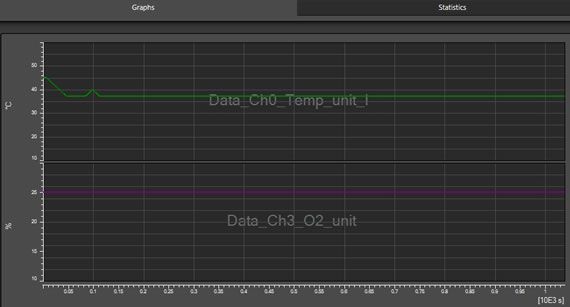
Full climate control of your experiment
All environmental data is saved with the experiment and can be reviewed subsequently. Know when changes in temperature, CO2 or O2 conditions occur that might affect your experimental results.
|
|
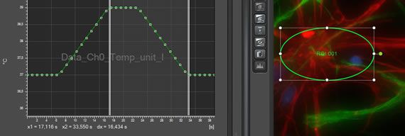
Run temperature profiles
In combination with LAS X Live Data Mode, macros can be generated to run temperature profiles. Acquisition can be set to start once a predefined temperature is reached. During the waiting time, a message displays the difference between nominal and actual environmental parameters. Alternatively, you can define that acquisition commences during ramping up of the temperature.
|
|
|
|
Get a warning when parameters are out of range
All environmental parameters are set within one interface. Activate range control to receive a warning when the upper or lower limits are exceeded. If needed, the experiment can be programmed to stop automatically once a limit is passed.
|
|
Don´t miss anything! With the LAS X Mobile Connection module you can stay connected to your experiment anywhere at any time. Display the imaging conditions on your remote device and review your experiment or download a specific image in full resolution and send it to a colleague. Start, pause, or stop experiments, or capture an image to identify the best time for starting your experiment remotely. |
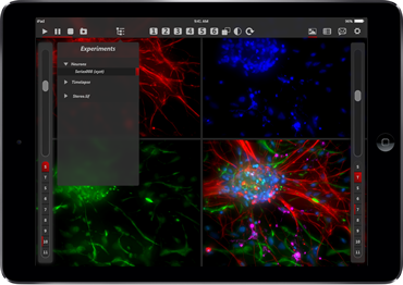
Connect to your experiment anywhere at any time
Connect to your acquisition station via web client or mobile device and monitor the course of your experiment, review the whole experiment or open any image series available in the experiment tree.
|
See imaging conditions at a glance
Review the channel settings defined for your experiment such as exposure time, gain, and filter cubes or take a look at environmental conditions like temperature, CO2 or O2 concentration (requires LAS X Environmental Control module).
|
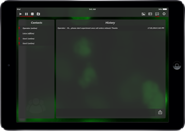
Chat and send messages to colleagues
Multiple remote users can be connected with the system at the same time. Chat with connected colleagues, send emails to other colleagues or send messages to the user at the acquisition station.
|
||||
 |
|
● Workflow oriented design for maximum ease of use, enabling even novices to become productive quickly
super-resolution systems - greatly reducing training time.
parfocality and parcentricity within LAS X. No need to exit the imaging software for reconfiguration and fine tuning.
during start up. User settings include all acquisition parameters, user specific configuration settings and layout of interface. |
● Full control of the microscope hardware while intelligent automation streamlines user interaction..
to future experiments for maximal reproducibility..
Operation Mode and design your own layout of the user interface. Display the menus where you need them..
between different acquisition configurations proposed by the system. Just enter the dyes of your sample and the confocal system will offer a set of possible system configurations along with their expected yield and cross talk.
|
|
|
Zebrafish Embryo
|
Zebrafish Embryo
|
Drosophila Adult
|
||||
Hiện tại chưa có ý kiến đánh giá nào về sản phẩm. Hãy là người đầu tiên chia sẻ cảm nhận của bạn.