|
|
All-Purpose Super-Sensitivity
The Hybrid Detector
|
|
Your Road to Super-Sensitivity
● Integrated into the Leica SP detector module for gapless multi-spectral imaging ● Superior sensitivity allows decreased light dosage ● Ideally suited for high-speed imaging ● Quantitative through single photon counting ● Descanned or non-descanned detection ● Modular concept of Leica TCS SP8 allows up to 4 HyDs ● NEW! Leica HyD SMD – the universal detector for FCS , FLIM, FLCS and super-sensitive imaging
|
|
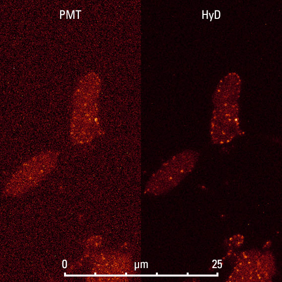
Neuromuscular junction in Drosophila melanogaster labeled with Bruchpilot::mStrawberry. The background of the PMT image is blurred by residual noise amplified by the maximum projection, while the HyD image is devoid of noise.
|
Low Dark Noise – High Contrast
Sensitive live specimens need to be imaged under low light conditions in high gain situations. Low noise can become the decisive advantage when it comes to recognizing weakly stained detail. Low dark noise is necessary for maximum signal efficiency, especially when photons are accumulated for more information. Otherwise, noise will pile up in the background of the image.
The Leica HyD offers superior signal-to-noise ratio to help render the finest details from any specimen – even tricky ones, such as highly scattering tissue slices. By reducing dark noise, the Leica HyD automatically improves image contrast. You obtain more information content, and the images are immediately publication-ready without any need for image processing. |
|
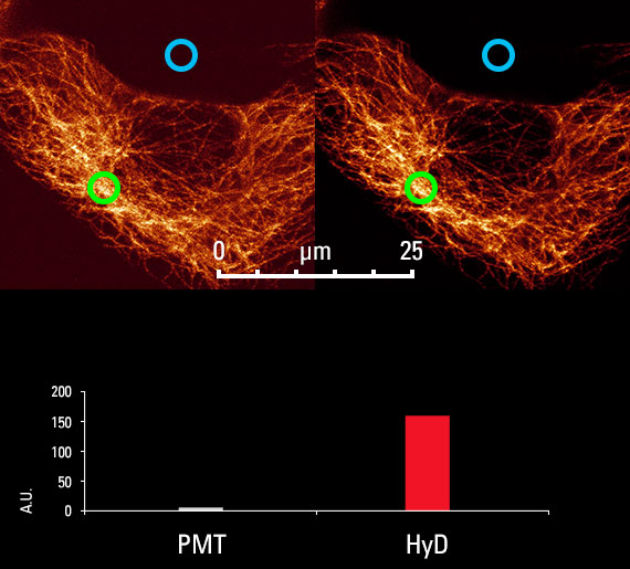
Leica HyD strongly improves contrast in comparison to PMTs
Sample: Tubulin
The contrast ratio was plotted as the ratio of mean intensity in the darkest region (blue) and the brightest region (green circle). |
Contrast of PMT vs. HyD
|
Three- to four-cell stage of Caenorhabditis elegans labeled by EGFP-tubulin. Courtesy of Prof. Pierre Gönczy, École Polytechnique Fédérale Lausanne, Switzerland.
|
High Speed Imaging with High Fidelity
Monitoring developmental processes involves collecting a series of images over time. In order to unravel the spatio-temporal formation of structure, one needs to find the right balance between acquisition speed and clarity. Along with Leica Microsystems’ pioneering tandem scanner, the Leica HyD offers unprecedented image quality. The tandem scanner’s resonant line frequency (8 or 12 kHz) leaves plenty of room for averaging or accumulation as needed, while retaining a large field of view.
|
|
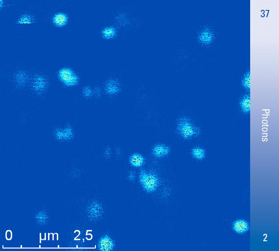
High Sensitivity for Single Molecules
Fixed single molecules represent the ultimate frontier of high-sensitivity imaging. When measuring such weak signals close to a reflective surface, the sensitivity, dark noise and efficiency of the beam splitting system are stretched to their limits. Note the half-moon shapes and horizontal lines in the image above, which show that molecules are blinking on and off. This reveals the single-molecule nature of the diffraction-limited spots.
|
|
Live yeast cells double-labeled with EGFP at both the nuclear envelope and the telomere. Courtesy of Prof. Susan M. Gasser, Friedrich Miescher Institute for Biomedical Research, Basel, Switzerland.
|
Increased Cell Viability for Sensitive Samplesy
Live cells can suffer from phototoxic effects as a result of imaging. While many of the underlying mechanisms are well understood, the effects of phototoxicity can be hard to pin down in the biological system being studied.
High sensitivity directly translates into reduced light delivered to the specimen and less bleaching. Even delicate systems such as yeast are detected by the Leica HyD – at full confocal resolution.
|
|
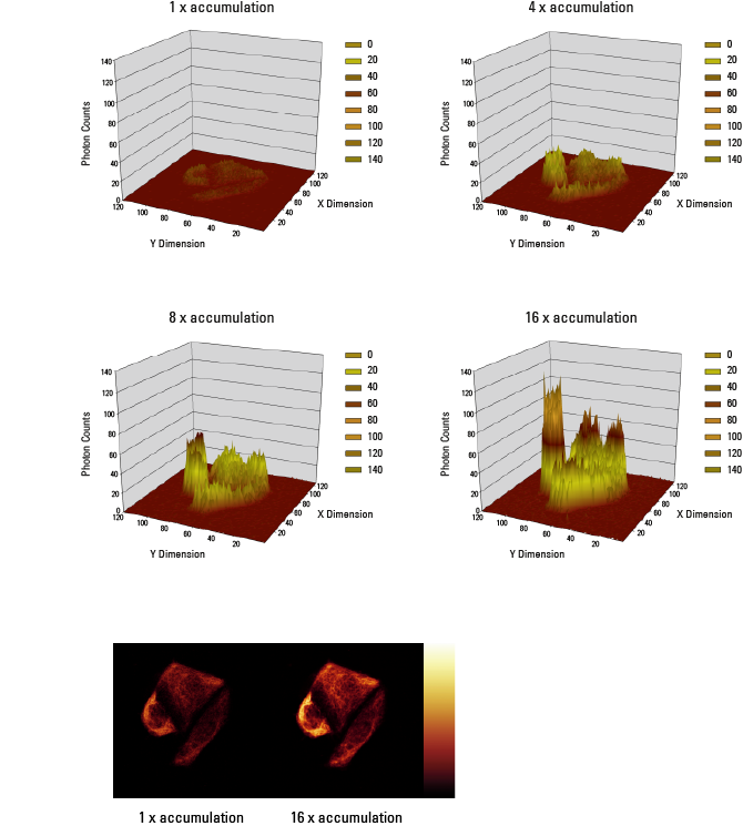
Maximum Dynamic Resolution by Photon Counting
Due to the very low background noise of our hybrid detectors, photon counting allows as much information to be accumulated as needed for any statistical analysis. In photon counting each pixel behaves like a bucket, which can be filled with photons. The longer one counts, the more photons are collected. The higher bit-depth modes available, 12 bit and 16 bit, represent very large buckets: in 12 bit mode, one can fill 4096, in 16 bit 65356 photons into one pixel. Thus, an enormous dynamic range with very low statistical per-pixel variance is available. The photon numbers are displayed via a look-up-table (LUT) on the screen. In this case the colors have a physical equivalent: photons.
|
Photon counting allows as much information to be accumulated as needed for statistical analysis.
|
Fast and easy NDD image acquisistion. Zebrafish embryo: lateral Line (GFP), neurons (DsRed), muscles (SHG), nuclei (BFP). Courtesy of Lionel Newton, EMBL Heidelberg (Gilmour lab).
|
Superior Sensitivity for Deep Tissue Imaging
Multiphoton microscopy has special requirements for signal detection, as the emitted light comes from deep tissue sections and is backscattered from surrounding structures.
To improve the efficiency of light collection, the detectors are placed as closely as possible to the source of emission (non-descanned detection, NDD).
A hybrid detector in RLD position gives you superior sensitivity for brighter images. Lower excitation power is needed that ensures less specimen damage, while a superior signal-to-noise ratio shows more details from deeper tissue sections.
The new QUAD module couples up to four hybrid detectors in RLD position, which gives you highest flexibility for multicolor deep tissue experiments. Due to its modular set-up you can choose as much super-sensitivity as you need for fast, easy non-descanned multicolor acquisition. |
|
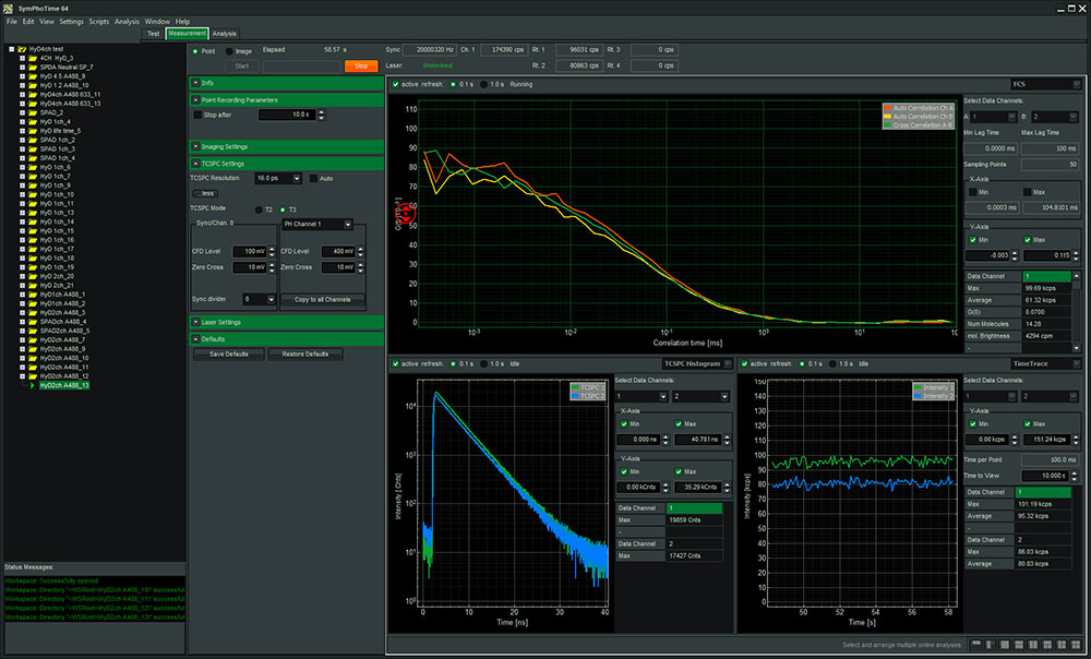
Leica HyD SMD – Universal Detector for FCS, FLIM, FLCS and Super-Sensitive Imaging
Analytical sensitivity is a prerequisite for reliable detection of single molecules. Leica Microsystems has developed a special hybrid detector with superior characteristics for all SMD (single molecule detection) methods fully integrated in the SP detection systems. An active cooling system comprised of built-in Peltier cooling and additional external cooling reduces the specified dark noise of the Leica HyD SMD, resulting in the highest SMD data quality.
● Virtually no detector after-pulsing allows acquisistion of precise diffusion data |
The flat FCS curve (top) shows after-pulsing and allows precise measurement of concentrations.
|
|
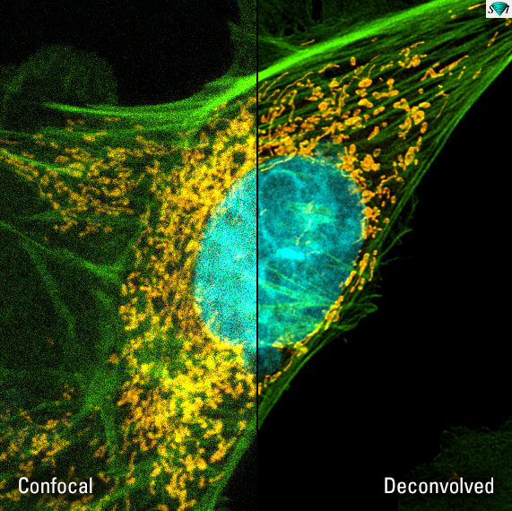
Applying deconvolution delivers detail-rich images with high contrast. MitoTracker® Red CMXRos (mitochondria, yellow), Alexa Fluor® 488 (F-actin, green), DAPI (nuclei, turquois). FluoCells® Prepared Slide #1, LifeTechnologies.
|
High Resolution Imaging with HyD and Huygens
The Leica HyD delivers crisp image detail with deep contrast. However, for deep-digging research tasks you might need to get more details from your confocal experiments. By combining our super-sensitive hybrid detectors and trusted mathematical Huygens deconvolution from the technology leader SVI, the Leica TCS SP8 high-resolution imaging system easily doubles the resolution of your confocal microscope and delivers crisp multicolor images that convey every detail at high fidelity. |
|
Hybrid Detector Technology – The Best of Two Worlds
Photodetectors translate light into electric signal, which makes them a critical part of the recording process. The Leica HyD combines the best characteristics of the classic PMT with the highly sensitive avalanche photodiodes (APDs). This results in super-sensitivity and large dynamic range combined with rapid detection speed and low dark noise, making them the ideal detectors for all samples.
Leica HyD photodetectors combine functional elements used in PMTs and APDs. The HyD’s photon detection is very efficient as there is virtually no loss of electrons. The dark noise level is very low, which gives an efficient recovery of the signal. The avalanche element allows immediate response and a very sharp electrical pulse. Photon counting is possible even at high intensities.
Find more information on HyD function and photon counting in our online tutorial. |
|
Hiện tại chưa có ý kiến đánh giá nào về sản phẩm. Hãy là người đầu tiên chia sẻ cảm nhận của bạn.


 Liên hệ với chúng tôi
 info@redstarvietnam.com